Advances in Stem Cell and Regenerative Therapies in Autoimmune and Metabolic Diseases: Implications for Healthy Longevity
Jonathan R. T. Lakey1, 2,*, Michael Alexander1, Mike K. S. Chan2, 4, Yuriy Nalapko2, 4, Michelle B. F. Wong2, 4 and Desiree C. T. Cox2, 3, 4
1University of California Irvine, Irvine, CA 92868, USA
2BioPep, 4621 Technology Drive Golden, CO 80403, USA
3Affiliate member, Graduate Faculty, School of Graduate Studies, Rutgers, The State University of New Jersey, Newark, NJ 07107, USA
4European Wellness International, Klosterstrasse 205a, Edenkoben, 67480, Germany
E-mail: jrtlakey@gmail.com; michaela@uci.edu; mtks3333@gmail.com; wbf6666@gmail.com; dr.desireecox@gmail.com
*Corresponding Author
Received 28 December 2023; Accepted 14 February 2024; Publication 20 April 2024
Type 1 diabetes (T1D) is an important autoimmune disease characterized by the destruction of insulin-secreting -cells in pancreatic islets of Langerhans resulting in hyperglycemia. More than 1.3 people in United States are suffering from T1D, and current treatments have failed to offer a lasting cure. T1D management requires lifelong daily injections of exogenous insulin to control blood sugar levels and secondary complications of this progressive disease, unless receiving pancreas or islet transplant with its associated risks from immunosuppression.
Peptide therapy aims to alter the response of cells by inducing or reducing cellular responses appropriate to the type of peptides used. The aim of this study was to investigate the protective effect on autoimmunity of Mito Organelle (MO) Peptides on the function and survival of peptides on pancreatic -cells in non-obese diabetes (NOD) mice model. MO peptide product (Biopep Inc) is a mixture of organ-specific cellular extracts that were extracted from organ specific cells, homogenized, filtered, and sterilized.
MO Peptides administered twice-weekly (IM) over 17-week period in NOD mice model were able to prevent the progression of autoimmune-mediated –cell destruction and the onset of hyperglycemia (blood glucose 300 mg dL) as compared to the saline treated control mice.
These studies will help understand the mechanisms of immunological protection in T1D and may serve as a model for other autoimmune disorders with peptides.
Keywords: Autoimmunity, diabetes, stem cell, peptides.
Type 1 Diabetes (T1D) affects around 1.3 million individuals in the United States, with the number expected to rise to 5 million people by 2050 [1]. One in 4 of US health care dollars spent on diabetes-related care, amounting to $237 billion in 2017 [2]. T1D is a chronic autoimmune disease that results from the destruction of the insulin-producing -cells in pancreatic islets of Langerhans [1–3]. Insulin is an essential hormone for regulation of blood glucose, by inducing transportation of glucose into cells and for metabolism by mitochondria. A deficiency in the production of insulin can lead to hyperglycemia, which when left untreated, can lead to long-term complications such as kidney disease, cardiovascular disease, peripheral arterial disease, stroke, and other diabetic-related complications. At present, there is no cure for T1D, and current treatments focus on the management of blood glucose levels through the lifetime administration of exogenous insulin [1–3].
These reports, as well as the understanding of immune cells involvement against endogenous or exogenous islets, led us to seek therapies based on reprogramming patients’ immune response. Among the long list of immune cells that can be targeted, macrophage’s role in T1D development has a high potential [3]. Autoimmune T1D follows a cascade of events that led to pancreatic -cell death: upregulation of pro-inflammatory cytokines, production of reactive oxygen species (ROS), and enhanced T-cell infiltration towards the pancreas. During acute inflammation, macrophages are the first cells to accumulate at one side of the pancreatic islet at an early stage (2–3 weeks of age) [4].
Peptide therapy aims to either induce peptide production or reinstate normal signaling patterns by renewing the strength of the signals received by cells [5–8]. Due to the differences in peptides produced between different tissue types, this therapy utilizes organ-specific extracts to target aging or diseased tissue with the goal of revitalizing normal peptidergic signaling in these regions. Protecting endogenous islets in T1D through the modulation of the immune system has been proposed [8]. Modulating the anti-islet T-cell response with antigen-specific peptide immunotherapy may offer the potential for preventing further tissue destruction and restoring immune homeostasis. Peptide immunotherapy offers the potential to prevent the immune-mediated response on the -cells and opens the possibility for -cell regeneration or replacement therapies to become more effective and long-lasting.
T1D represents an ideal autoimmune disease for testing peptide therapy because of the extensive knowledge of the etiology and pathology of the disease, and the availability of the non-obese diabetic (NOD) mouse model that parallels the disease course in humans [9]. NOD mouse model is widely used for T1D research due to its natural development of diabetogenic T-cells that lead to the destruction of the -cells of the pancreatic islets of Langerhans and result in insulin deficiency [9]. As early as 3 weeks of age, the innate immune cells of NOD mice infiltrate the islets of the pancreas and trigger the response of the adaptive immune system. As the NOD mice age, autoreactive T-cells that recognize specific diabetes-related autoantigens begin to develop within the thymus and escape negative selection into the periphery [10]. Once in the periphery, these T-cells fail to be selected against, and instead become activated, leading to the destruction of the insulin-producing -cells in the pancreatic islets. NOD mice also exhibit similar symptoms to diabetic humans such as glycosuria, polydipsia, weight loss, and polyuria [10]. A prior study [11] found that the administration of mitochondrial-encoded peptides in NOD mice could modulate T-cells and induce changes in the function and phenotype of the immune cells. These results indicated a significant decrease in the incidence of diabetes in NOD mice and the reduction of T-cell activation, supporting the notion that peptide therapy may ameliorate the progression or prevent the onset of T1D in humans.
European Wellness (EW) and the BioPep Research Group have developed a peptide therapy product made of organ-specific cellular extracts and peptide molecules that are intended for therapeutic use in both animals and humans [12–16]. EW and BioPep manufacture two distinct peptide products, Mito Organelles (MO) peptides and Nano Organo Peptides (NOP), which are produced through a proprietary parallel-extraction process from mammalian precursor stem cells and rabbits bred in closed colonies under good manufacturing practices conditions. MO peptides are biologically extracted mixtures of cellular peptides that have predominantly mitochondria-specific functions [15, 16]. Unlike NOPs, which are under 10 kDa in size due to their extraction process, MOs are larger in size and have predominantly mitochondria-specific functions allowing for a more pronounced revitalization of mitochondrial deficits. MO peptides are organ-specific extracts that aim to revitalize and rejuvenate mitochondrial activity, thereby regenerating diseased or aging tissue. These mitochondria-derived peptides (MDPs) are widely distributed through various tissues and have been shown to play cryoprotective roles through maintaining cell viability and mitochondrial function under both pathological and normal conditions [15, 16]. This study seeks to determine whether the administration of stem-cell derived MO peptides twice-weekly to NOD mice through intramuscular injections over 17 weeks delays or prevents the onset of the destruction of the insulin-secreting -cells in pancreatic islets of Langerhans. Specifically, the stem-cell derived MO peptides utilized in this study will be obtained from thymus and pancreatic extracts to target the regions of the -cells and T-cell maturation [17, 18]. It is hypothesized mice administered with MO peptides will have better maintained blood glucose levels throughout the study and be less susceptible to the development of T1D as compared to control. Evidence of a positive effect on survival and function of MO peptides on -cells would provide important evidence for the protective effect of MO peptides on delaying hyperglycemia and diabetic onset.
This study was conducted under approved Institutional Animal Care and Use Committee at University of California Irvine, protocol # AUP-17-241.
A total of 12 female NOD mice (Charles River, San Diego, CA) were purchased at an age of 6 weeks. Once acclimated to the facility, the mice were divided into MO peptide and saline injection groups. The selected concentration of MO peptides for administration was based on the human injection dose scaled down to the body weight of an average mouse (approximately 30–32 g). The regular human dose of MO peptides is 7 g/kg (0.035 mL/kg), and since each MO dose consists of approximately 500 g, the dilution for the mice was scaled accordingly. Per weekly dose, 100 L of MO peptide solution was diluted with 10 mL of saline to achieve the same concentration used in humans. Mice received either 90 L of the diluted MO peptide solution or 90 L of saline solution as a control administered subcutaneously twice a week. The first injection of MO peptide or saline solution was administered at week 1 and the last injection was administered at week 17.
Blood glucose measurements were obtained once a week in all mice using the tail capillary prick method. At week 1, blood sugar levels were recorded immediately before the administration of the first dose of peptide or saline. Therefore, week 1 blood glucose readings represent the NOD mice basal glucose level and week 2 values indicate changes in blood glucose a week after the first round of injection of MO peptide. For mice that had a blood glucose concentration 300 mg/dL, a follow up measurement was taken 2 days later. If the blood glucose concentration remained 300 mg/dL, the mouse was deemed diabetic and sacrificed. An average of the two measurements was then used for the results in that given week. Mice were monitored for a total of 17 weeks unless they had three consecutive non fasting blood glucose readings from capillary blood collected from the tail vein and read using a commercial blood glucose meter.
Data analysis and graph construction were performed in Microsoft Excel®. A two-sample t-test method was selected to compare the weekly blood glucose levels of the MO peptide and saline mice. Statistical testing was performed to compare blood sugars between the MO peptide treat and the saline control group twice a week until the mice reached the age of 17 weeks.
Statistical testing was also performed to compare weekly blood sugar levels between the NOD saline and NOD MO treated group of mice to ensure groups were representative of each other using standard t test. A probability of 0.05 was considered statistically significant.
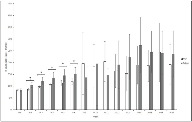
Figure 1 The change in the mean blood glucose concentration (mg/dL) of NOD mice in the MO peptide (n 6) and control group (n 6) over 17 weeks. Week 1 values represent the mean blood glucose level before the first injection of MO peptide or saline. Following the sacrifice of mice that were deemed diabetic (two consecutive readings 300 mg/dL), a blood glucose concentration of 300 mg/dL was substituted for all subsequent weeks to provide a more accurate comparison of groups. Statistical analysis was performed after the first injection to compare the blood glucose concentration between the MO peptide and saline group each week. * Represents a significant difference (p 0.05). Error bars represent mean SEM.
Baseline measurements of non-fasting blood glucose concentration in adult NOD MO peptide mice and saline control mice were collected at week 1 of the study (Figure 1). Initially, we found no statistical difference in the blood glucose concentration was found between the groups (p 0.532). The blood glucose level of the MO peptide group was significantly lower than the saline group from week 2 to week 6 (p 0.05, paired t test). Blood sugar monitoring resumed at week 9 and no significant difference was found in the blood glucose levels of the two groups on weeks 9 (p 0.327), 10 (p 0.702), 11 (p 0.867), 12 (p 0.508), 13 (p 0.110), 14 (p 0.253), 15 (p 0.376), 16 (p 0.329), and 17 (p 0.257). Although the blood glucose concentration of MO peptide and saline groups were not significantly different from week 10 to 13, the mean blood sugar in the MO peptide group began to drop on week 11 until the conclusion of the experiment at week 17.
Table 1 Mean weekly blood glucose concentrations (mg/dl) between NOD MO peptide mice and controls. Mean weekly blood glucose concentrations (mg/dl) between NOD MO peptide mice and controls. Mice that were deemed diabetic (two consecutive readings 300 mg/dL) were not included in the mean weekly blood glucose concentration (mg/dL) calculation following their sacrifice
| W1 | W2 | W3 | W4 | W5 | W6 | W9 | W10 | W11 | W12 | W13 | W14 | W15 | W16 | W17 | |
| MO Peptide (mg/dL) | 85 | 88 | 98 | 107 | 114 | 120 | 196 | 161 | 186 | 139 | 125 | 168 | 164 | 233 | 138 |
| Saline Control (mg/dL) | 83 | 105 | 121 | 135 | 145 | 152 | 137 | 196 | 197 | 170 | 207 | 251 | 133 | 123 | 113 |
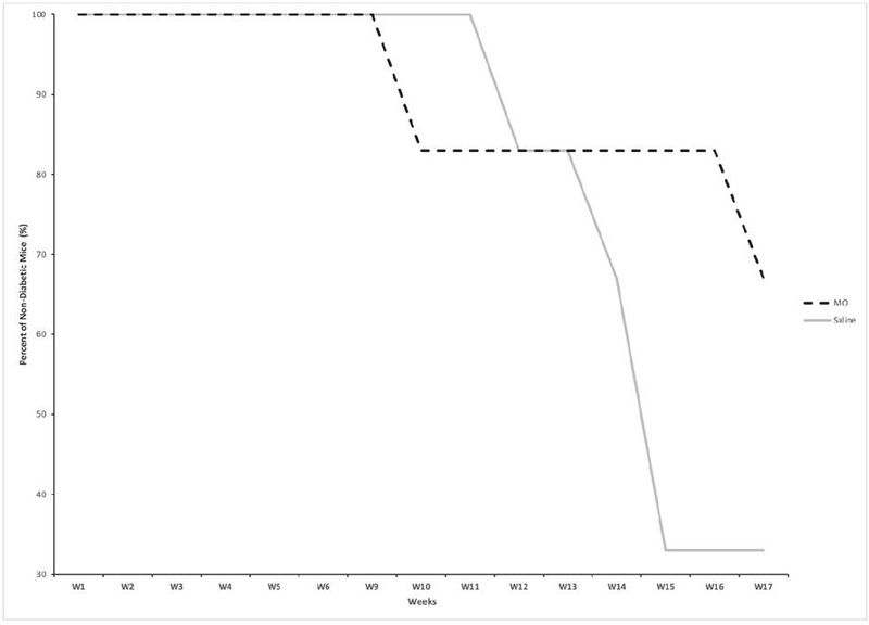
Figure 2 The change in percentage of non-diabetic NOD mice in the MO peptide (n 6) and control groups (n 6) over the course of 17 weeks. All mice that had two consecutive blood glucose readings 300 mg/dL were sacrificed and were no longer included in data recordings.
All mice in both MO peptide and saline groups were non-diabetic up until week 9 (Figure 2). Four out of the six MO treated mice remained non-diabetic at the end of the study, while only two of the six control mice remained non-diabetic. Within both groups, mice which developed diabetes (blood glucose 300 mg/dL) during the study were sacrificed and blood glucose measurements were continued only for non-diabetic mice. Although the proportion of non-diabetic mice in the MO group showed an initial decrease at week 9 relative to the control mice, the control mice showed an overall greater decrease in the proportion of non-diabetic mice by week 17 of the study. There was a 33% greater non-diabetic population in the MO peptide treated mice versus the saline treated mice by the end of week 17.
The administration of exogenous peptides, known as peptide therapy, aims to either induce peptide production or reinstate normal signaling patterns by renewing the strength of the signals received by cells. Due to the differences in peptides produced between different tissue types, peptide therapy utilizes organ-specific extracts to target aging or diseased tissue with the goal of revitalizing normal peptidergic signaling in these cells and improving overall health. Prior research has demonstrated the effectiveness of peptide therapy in decreasing the incidence of diabetes in NOD mice and reducing T-cell activation [11, 15].
Our study sought to investigate whether the administration of MO peptides over the course of 17 weeks would affect the development of autoimmune diabetes in the NOD mouse model. The MO peptides are mitochondrial derived organ specific peptides produced by European Wellness and BioPep Research Group through a proprietary parallel-extraction process.
All mice in both MO peptide and control groups were non-diabetic up until week 9 (Figure 2). Four out of the six MO treated mice remained non-diabetic at the end of the study while only two of the six controls mice remained non-diabetic. Although the percent of non-diabetic mice in the MO group had an initial decrease at week 9 relative to the saline mice, the mice in the saline group showed an overall greater decrease in percent of non-diabetics by week 17 of the study. Results showed a 33% greater non-diabetic population in the MO peptide treated mice versus the saline treated mice by the end of week 17. At the conclusion of the study on week 17, all remaining NOD mice were euthanized.
Together, these results indicate that an underlying change to the signaling and physiology of the NOD mice occurred. These changes may have protected from the destruction of the -pancreatic cells of Langerhans and resulted in the delayed onset or possible prevention of diabetes in the treatment group. However, it’s important to note the small sample size and relatively short study time. Further research is necessary in longer and larger studies, as well as to determine the exact mechanism/s by which this protective effect occurred.
This pilot study demonstrates the beneficial effects of intramuscular delivery of stem cell derived peptides in delaying the onset of autoimmune diabetes in NOD mice. It provides important preliminary data that suggests that MO peptides may assist in delaying the onset or preventing T1D and represents an exciting therapeutic option to further investigate. Future studies will focus on timing of delivery of organ specific stem cell peptides and further investigate the dose of peptide and duration of effect.
This project was funded by BioPep Inc. The authors also recognize and appreciate the support from the Sue and Bill Gross Hall Stem Cell Center, a California Institute of Regenerative Medicine (CIRM) at the University of California, Irvine.
This article is part of the journal’s collaboration with The HEALinc, a global future health research and development organization, Bahamas. The subject of this particular article was presented at the 2023 HEALinc Future Health Summit.
JL and MA analyzed data and wrote the manuscript; MC, MW, and DC reviewed and edited the manuscript; DC secured funding for the manuscript.
[1] American Diabetes Association, ‘Economic Costs of Diabetes in the U.S. in 2017’, Diabetes Care, pp. 917–928, May, 2018.
[2] G. Imperatore, E.J. Mayer-Davis, T.J. Orchard, et al., ‘Prevalence and Incidence of Type 1 Diabetes Among Children and Adults in the United States and Comparison with Non-U.S. Countries’, In: Diabetes in America, 3rd edition, Chapter 2, Bethesda, Aug., 2018.
[3] M. Wallberg, A. Cooke, ‘Immune mechanisms in type 1 diabetes’, Trends in Immunology, pp. 583–591, Dec., 2013.
[4] K.C. Navegantes, et al., ‘Immune modulation of some autoimmune diseases: the critical role of macrophages and neutrophils in the innate and adaptive immunity’, J Transl Med, pp. 36, Feb, 2017.
[5] D. Klokol, L. Nallenthiran, M.K.S. Chan, et al., ‘Cell therapy as the main stratagem of anti-aging and regenerative medicine’, Europ J Pharm Med Res, pp. 295–299, May, 2019.
[6] A.C. Lee, J.L. Harris, K.K. Khanna, et al., ‘A comprehensive review on current advances in peptide drug development and design’, Int J Mol Sci, pp. 2383, May, 2019.
[7] B.G. De La Torre, F. Albericio, ‘Peptide therapeutics 2.0’, Molecules, pp. 2293, May, 2020.
[8] E.L. Smith, M. Peakman, ‘Peptide immunotherapy for type 1 diabetes-clinical advances’, Frontiers in Immunol, pp. 392, Feb., 2018.
[9] A. Jansen, F. Homo-Delarche, H. Hooijkaas, et al., ‘Immunohistochemical characterization of monocytes-macrophages and dendritic cells involved in the initiation of the insulitis and beta-cell destruction in NOD mice’, Diabetes, pp. 667–675, May, 1994.
[10] E.H. Leiter, M. Prochazka, D.L. Coleman, ‘The non-obese diabetic (NOD) mouse’, The American J of Pathol, pp. 380-383, Aug., 1987.
[11] B.S. Kong, S.H. Min, C. Lee, et al., ‘Mitochondrial-encoded Mots-C prevents pancreatic islet destruction in autoimmune diabetes’, Cell Reports, pp. 109447, Jul., 2021.
[12] D. Klokol, M.K.S. Chan, M.B.F. Wong, ‘European wellness – the evidenced rationale behind the biological medicine: ad astra per aspera’, J Pharm Biomed Sci, pp. 19–22, 2017.
[13] R. Moya, M.K.S. Chan, M.B.F. Wong, et al., ‘Active specific immunotherapy (asi) in cancer treatment: five case reports’, J Cancer Sci Ther, pp. 256–259, 2019.
[14] D. Klokol, L. Nallenthiran, M.K.S. Chan, et al., ‘Cell therapy as the main stratagem of anti-aging and regenerative medicine’, Europ J Pharm Med Res, pp. 295–299, Jun., 2019.
[15] C. Lee, J. Zeng, B.G. Drew, et al., ‘The mitochondrial-derived peptide mots-c promotes metabolic homeostasis and reduces obesity and insulin resistance’, Cell metabolism, pp. 443–454, Mar, 2015.
[16] D. Wu, E. Kampmann, G. Qian, ‘Novel insights into the role of mitochondria-derived peptides in myocardial infarction’, Frontiers in Physiology, pp. 750177, Oct., 2021.
[17] P. Thapa, D.L. Farber, ‘The role of the thymus in the immune response’, Thoracic Surgery Clinics, pp. 123–131, May, 2019.
[18] B. Saberzadeh-Ardestani, R. Karamzadeh, M. Basiri, ‘Type 1 diabetes mellitus: cellular and molecular pathophysiology at a glance’, Cell Journal, pp. 294–301, May, 2018.
International Journal of Translational Science, Vol. 1, 115–126.
doi: 10.13052/ijts2246-8765.2024.023
© 2024 River Publishers