Mechanisms, Models, and Clinical Applications of Cell Membrane Electroporation
Daniele Marazzi1, †, Felicia Carotenuto1, †, Federica Trovalusci2, Patrizia Caruccio2 and Paolo Di Nardo1, 3,*
1Department of Clinical Sciences and Translational Medicine, School of Medicine, University of Rome “Tor Vergata” Rome, 00133, Italy
2Department of Enterprise Engineering “Mario Lucertini”, University of Rome “Tor Vergata”, Rome, 00133, Italy
3Interdepartmental Center for Regenerative Medicine (CIMER), University of Rome “Tor Vergata”, Rome, 00133, Italy
E-mail: dinardo@uniroma2.it
*Corresponding Author
†Equally contributed
Received 18 November 2024; Accepted 01 December 2024
Abstract
Electroporation is an essential biophysical process that involves the use of pulsed electric fields to temporarily increase the permeability of cell membranes. A comprehensive overview of the main clinical and biomedical applications of cellular electroporation is provided here with a particular focus on cancer therapy, genetics, and drug delivery. The review concentrates on the characterization of membrane stresses caused by electroporation and their impact on cell membrane structure and dynamics. It analyses the relationship between applied electric fields, cell geometry, and membrane composition with the aim of developing mathematical models to simulate cell geometries and the electroporation process. Furthermore, it identifies both the beneficial effects and potential complications of the electroporation treatment, as well as the various mathematical models that have been developed to simulate the effects of such treatments. The use of sophisticated simulation algorithms enables an in-depth investigation of the intricate relationship between electrical parameters and cellular responses, facilitating a comprehensive assessment of the efficacy and safety of the electroporation procedures.
Keywords: Electroporation, nuclear and cell membrane permeability, drug delivery, mathematical modeling, cancer therapy.
Introduction
The cell membrane, which is composed of a complex lipid–protein structure, delimits the outer boundary of cells and plays a fundamental role in cellular homeostasis [1, 2]. In addition to its function as a physical barrier, it acts as a biophysically and biochemically regulated interface, allowing the selective exchange of molecules between the cytoplasm and extracellular environment [3]. As observed by Catterall (2000), cell membrane potential is mainly modulated by the ion exchange of potassium (K+), sodium (Na+), and chloride (Cl-) through ion channels that are central to the maintenance of ion homeostasis and cellular signal transduction [4]. Through a wide range of transporter proteins, ion channels, and receptors, the cell membrane carefully regulates the flow of ions and molecules, facilitating the uptake of vital nutrients and removal of waste products [5, 6].
In addition to the transport of nutrients, the cell membrane also participates in the translation of signals through biochemical mechanisms and electrical stimuli. External messages are interpreted by specific receptors on the membrane, triggering intracellular signals that affect vital processes such as proliferation, differentiation, and immune response [7]. This interaction between the cell membrane and surrounding environment is critical for maintaining physiological integrity and regulating cellular and tissue functions.
Given the critical role of the cell membrane in regulating ion exchange and maintaining cellular homeostasis, techniques that can temporarily alter membrane permeability have significant biomedical applications [8]. Among the different methods that allow the modification of the cell membrane, electroporation has been widely used as a non-invasive technique to increase cell membrane permeability [9]. The electroporation procedure consists of applying a pulsed electric field of a specific intensity and duration to the cell membrane, stimulating the formation of transient pores that promote ion exchange between the cytoplasm and the surrounding environment.
To optimize the electroporation process, numerous studies have been conducted aimed at developing mathematical and simulation models capable of reproducing the phenomenon [63]. These models are essential for understanding the interactions between the electric fields and cell membranes, as well as for predicting how changes in electrophysical parameters may affect treatment effectiveness.
Over the years, several models have emerged, each with a different degree of complexity and accuracy. Some of these models are based on differential equations that describe the dynamic behavior of cells under electrical stress, while others use computer simulation-based approaches to visualize processes at the microscopic level. These models made it possible to identify the optimal methodologies for the application of the electroporation technique, highlighting key parameters such as electric field strength, pulse duration and lipid composition of the cell membrane. This computational optimization made it possible to maximize the efficiency of the transfer of substances within cells, while reducing the side effects associated with, e.g., cytotoxicity [65].
In terms of simulating the electroporation phenomenon, a specific finite element method (FEM) software is utilized, which, through appropriate initial parameters, enables the simulation of the behavior of biological material [11]. However, the simulation and implementation of models for nuclear electroporation processes present several challenges. These challenges stem from the fact that it is not possible to perfectly predict and simulate all aspects of the electroporation process, as the simulation of physical and chemical processes involves a calibration phase that is linked to the quality of stochastic data from previously conducted studies [67, 68]. Additionally, the simulation of the process allows for the development of customized protocols based on the type of cell and the molecules to be introduced, ultimately resulting in a prediction of transfection efficiency [12].
In this review, an in-depth analysis of the main aspects of electroporation in the biomedical context will be conducted, with particular emphasis on the critical issues associated with this technology. These critical issues, which include the management of cell permeability, the control of cytotoxicity, and the variability in individual responses to treatment, will require careful and systematic evaluation. It is essential to address these challenges to ensure not only the safety, but also the efficacy of clinical applications of electroporation, thus allowing therapeutic protocols to be optimized and patient outcomes to be improved. The latest clinical applications of electroporation will be examined, including innovations in cancer treatment and gene therapy. Emerging issues will be discussed, such as the need to optimize treatment protocols and associated technologies, as well as the strategies to improve the integration of pharmacological agents, to maximize therapeutic efficacy.
Finally, the main mathematical models that have contributed to optimizing the clinical and biological processes of electroporation will be analyzed. These models provide a theoretical basis for understanding the dynamics of cell permeabilization and offer predictive tools to improve treatment protocols. The importance of these models in guiding future research and facilitating the clinical application of the technique will be discussed, helping to make electroporation an increasingly effective and safe therapeutic strategy.
Electroporation Mechanism
Electroporation is a biophysical method that employs an electric field to the cell membrane, triggering a range of biophysical and biochemical reactions. These reactions are directly affected by the cell’s transmembrane tension, which can be explained using various mechanisms and relevant parameters [9]. A crucial factor in this technique is the transmembrane potential, primarily influenced by the activity of sodium (Na+, 139.1 0.6 mmol/l) [13] and potassium (K+, 140–150 mmol/l) [14] ion channels. This potential, also referred to as the membrane potential, represents the voltage differential across the cell membrane. It plays a vital role in numerous cellular processes, including maintaining cellular homeostasis, transmitting signals, and controlling ion movement.
The electroporation process begins with the application of an electric field, which will alter the distribution of charges on the surface of the cell membrane [15]. This differentiation of charges causes an increase in the trans-membrane voltage which, once a certain critical threshold is exceeded, will lead to the formation of pores [16]. These pores will be characterized by a certain temporary permeability, which will allow the passage of ions and/or molecules inside the cell [17]. Pore formation is mediated through several mechanisms, which are shown in Table 1 [18, 19].
Table 1 Mechanism of electroporation
| Mechanism of | |
| Electroporation | Description |
| Puncture | The energy of the electric field induces elongation and deformation of the membrane, generating pores that allow the passage of ions and small molecules. |
| Electro-osmotic | The electric field induces a movement of the solvent (i.e. water) through the membrane, contributing to both the formation of pores and the diffusion of molecules. |
| Ionic | Variation in membrane potentially affects the activity of ion channels, such as those for sodium and potassium, further affecting the ion balance and stability of the membrane during and after electroporation. |
In a cell’s resting state, the plasma membrane’s higher conductance to potassium compared to sodium leads to potassium ions flowing out of the cell, creating a negative membrane polarization known as the resting potential [10]. However, external stimuli, such as nerve impulses or hormonal signals, can swiftly activate sodium channels, causing a temporary change in the transmembrane potential called the action potential [11]. The membrane potential, which is the electric potential difference between the cell’s interior and exterior, is maintained at a stable value termed the “resting potential” in quiescent cells, typically ranging from 70 mV to 40 mV in neurons. This potential is essential for cellular function and homeostasis, providing energy for active substance transport and facilitating signal transmission [12].
Polarization is crucial for proper cell function. When an electric field is applied, it generates a new potential that overlaps with the cell membrane’s resting potential. If the voltage applied to the membrane produces a transmembrane electric field below the critical threshold value, the membrane responds elastically. This response preserves the membrane’s structural and functional integrity, characterized by temporary deformation without pore formation or permanent damage [13, 14]. Conversely, if the transmembrane potential surpasses a critical threshold, temporary pores form in the membrane structure. This phenomenon enables the entry of molecules or molecular complexes, such as therapeutic agents, into the cells [15].
Electroporation can manifest in two forms: reversible and irreversible [16]. In the reversible variant, the cell membrane pores can spontaneously seal after the electric field is removed, thus restoring the membrane’s structural integrity [17]. This method specifically targets cell membranes, leaving collagen structures and associated proteins unharmed, which offers a significant benefit for tissue healing due to the limited damage [18]. Furthermore, while less pronounced than conventional techniques, there are thermal advantages, provided the appropriate validity parameters are established [19].
Contemporary research has identified several crucial factors that affect pore formation and reversibility during electroporation [22]. These encompass transmembrane tension, pore density, average pore radius, and thermal regulation during electric-field application. Adhering to these factors is essential for ensuring the effectiveness and safety of electroporation in biomedical applications [23]. For instance, research by Pliquet et al. demonstrates that the dynamic membrane resistance undergoes a notable change when pulses ranging from 40 to 80 V are applied and stabilizes at resistance values between 50 and 100 [24].
Optimizing Electroporation Parameters
When applying electroporation to cells, it is crucial to accurately identify and adjust various parameters associated with the electrical impulse [34]. This process of parameter identification and adjustment is vital for achieving effective and controlled outcomes [35]. Several factors must be considered when applying electroporation to cells, as shown in Table 2.
Table 2 Cellular factors that influence electroporation
| Cell Parameter | Description |
| Type | Different cell types of exhibit variations in their susceptibility to electroporation, partly determined by differences in membrane lipid and protein composition. Therefore, it is important to adapt the parameters of the electrical impulse according to the type of cell being treated [20]. |
| Size | Cell size directly affects the distribution and strength of the applied electric field. Cells of different sizes require suitable electrical pulse parameters to ensure uniform electric field penetration and homogeneous membrane response [21]. |
| Orientation | The orientation of cells relative to the electric field can significantly affect the effectiveness of electroporation [22]. |
| Density | Cell density can affect the distribution of the electric field and the spread of therapeutic agents through the cells. The optimal density varies depending on the specific application and the characteristics of the cell sample [23]. |
The duration of electroporation-induced membrane permeability is modulated by factors such as the applied electric field strength, pulse duration, and the specific lipid composition of the plasma membrane. Recent studies indicate that, although electroporation induces a pronounced increase in membrane permeability, cellular membranes are capable of rapidly restoring their structural and functional integrity once the electric field is withdrawn [31]. This restoration is facilitated by intrinsic membrane repair mechanisms, including pore closure and the reorganization of integral membrane proteins, which collectively re-establish membrane barrier function on a microsecond timescale [32].
Several protocols have been developed that determine the optimal adhesion conditions for a wide range of applications. The protocols include a preliminary phase, in which the cells to be subjected to treatment (mammalian cells, plant cells, etc.), the type of substance to be introduced into the cell (plasmids of DNA, RNA, oligonucleotides, proteins, etc.), and the culture medium in which cell growth will take place will be identified [24]. Then there is a preparatory phase, in which the cell culture will be carried out and the substances that are to be introduced into the cell will be prepared. At the end of the preparatory phase, the cells will be mixed with the substance, and then inserted into appropriate cuvettes for electroporation [25]. Once everything has been prepared, the different parameters to be used for the experiment will be set on the electroporator, such as the current intensity, the duration of the pulse, the number of pulses, the time constant inherent in the discharge circuit, the type and concentration of the electroporation medium and the temperature. The above parameters vary according to the cell to be treated and, in the specific case of mammalian cells, which are reported in Table 3.
Table 3 Electroporation parameters for mammalian cells
| Parameters | Value |
| Electric field strength | 1500–2000 V/cm |
| Pulse duration | 5–20 ms |
| Number of pulses | 1–3 |
| 10–20 ms | |
| Electroporation medium | PBS or HBSS in appropriate concentrations |
| Temperature | Constant at 37 C (310 K, physiological temperature) |
The above parameters will require an optimization step, based on the specifications of the cells, the substance to be introduced, and the experimental conditions, to obtain better results in terms of cell transfer efficiency and survival [25].
At the end of the electrical stimulation phase, the cells undergoing treatment will be recovered, which must be incubated under appropriate conditions. Finally, there is a phase of analysis of the treatment, in which the efficiency of electroporation will be evaluated through specific methods, such as PCR, with which we can verify the transfection of the substance into the cells, the efficacy and cellular integrity.
Clinical Use of Electroporation
Electroporation, a multifaceted technique, finds applications in various biomedical and biological fields, such as targeted intracellular drug delivery, genetic therapy, cellular fusion, sterilization of tissues, and ablation [16]. A significant advancement in electroporation has been the incorporation of pharmacological agents into its protocols. This progress has significantly enhanced the effectiveness of cellular treatments, thereby increasing electroporation’s utility in biomedical applications [33]. The protocols for electroporation are meticulously designed to correspond with the particular aims and cellular properties of the targeted tissue, enabling precise adaptation to fulfil the needs of each experimental or clinical use [42, 43].
Electroporation-based drug delivery is a crucial medical therapy, particularly in the administration of anticancer agents. This method creates transient openings in cellular membranes, enhancing the uptake of specific drugs by cancer cells [37]. This targeted approach offers a selective treatment option that minimizes harm to adjacent tissues and reduces the adverse effects commonly associated with traditional therapies [44].
Alongside electroporation, other novel techniques such as electrochemotherapy are utilized in cancer treatment [45]. Electrochemotherapy integrates electrical stimulation with drug therapy to target cancer cells. This method has demonstrated effectiveness in selectively attacking cancerous cells whilst preserving healthy surrounding tissue.
The process of electrochemotherapy involves the application of electrical pulses in conjunction with the administration of drugs that are typically non-permeable or have low permeability across cell membranes. The electrical stimulation temporarily enhances membrane permeability, enabling cytotoxic drugs to enter cancer cells more readily [46]. The drugs employed in this protocol are generally cytotoxic agents that, despite their limited ability to traverse cell membranes independently, exhibit potent intracellular activity once inside the target cell. This strategy concentrates pharmacological agents within cancer cells, thereby compromising their viability and reducing tumor size [47].
A notable advantage of electrochemotherapy is its ability to selectively target and destroy cancerous cells while limiting damage to adjacent healthy tissue. This characteristic sets it apart from other treatments that may cause more extensive collateral damage. However, like any medical procedure, it is essential to carefully evaluate the risks and benefits of electrochemotherapy for each patient and cancer type, and to administer treatment under the supervision of experienced healthcare professionals [48].
The electroporation is effective if the cells have a good permeabilization, which is why the cells are subjected to an electric field. However, the electric field will need to be as low as possible to ensure the safety of the procedure. For this reason, appropriate electrodes must be carefully developed, and appropriate parameters identified. In particular, the following types of electrodes can be identified:
• Plate electrodes, used for surface treatments.
• Needle electrode, which allows localized treatment. Depending on the geometry of the needle, we can process different sizes of areas. In particular, the hexagonal needle electrode is an electrode that can treat large areas.
The above electrodes can be made of surgical steel (AISI 316L) or aluminum (the latter only for experimental purposes) [26].
Through several studies, conducted both in vitro and in vivo, it has been possible to define standard parameters for the delivery of electrical impulses. This involves 8 square wave pulses of 100 s with a voltage of about 1000 V, delivered at a frequency of 1 Hz or 5 KHz. However, there are conflicting experimental results in the literature regarding the efficacy of this therapy for these frequencies, as several articles report therapeutic efficacy even at lower frequencies and with higher efficiencies [27].
The comparison between high ionic conductivity and the permeability state of the cell membrane poses a fundamental challenge in the current scientific context, highlighting the limitations of existing mathematical models in adequately understanding these phenomena. This complexity is intrinsic to the intricate nature of the biophysical and molecular processes that govern cell membrane dynamics [28, 29].
Current models, such as those based on membrane potential, are configured as simplifications of the complex biological reality, treating the cell membrane as a single homogeneous entity. However, this view neglects the structural and functional variations present in the membrane, such as the presence of specialized lipid domains and the diversification in the distribution of ion channels and membrane transporters [30, 31].
Existing models tend to overestimate the duration of the conduction state compared to that experimentally observed in the permeability state. This discrepancy could be attributed to the lack of a detailed representation of the molecular mechanisms involved in the passage of solutes across the membrane, such as the formation of pores or transient ion channels [30, 32].
Further complications in the analysis of mathematical models arise from the additional complexity introduced by the lipid bilayer structure of the cell membrane, which is characterized by a wide range of molecular interactions and phase dynamics that can significantly affect membrane permeability and conductivity [33].
To address these challenges, it is necessary to develop more sophisticated mathematical models that integrate a detailed understanding of cell membrane structure and dynamics. Only through an in-depth analysis of such models will it be possible to obtain an accurate and predictive understanding of the behavior of the cell membrane in conduction and permeability states, with significant implications for biological and medical research [16, 33].
Nuclear Electroporation
An important aspect that has emerged in recent years is the ability to deliver electrical stimulation directly to the nucleus of a cell, a technique known as nuclear electroporation. This method enables biological materials, such as DNA, mRNA, or proteins, to be inserted directly into the nucleus of a cell, bypassing the outer cell membrane, and resulting in a more targeted therapy [34, 35]. Unlike traditional electroporation, nuclear electroporation involves the creation of pores in the nuclear membrane through precise electrical stimulation, ensuring a more efficient and targeted transfer of genetic material.
The electrical stimulation of the cell nucleus is a more complex process than that of the outer cell membrane due to the involvement of various physical and chemical processes within the cell [60]. Physical processes include the conduction of electricity through cellular tissues and the generation of electric fields that affect the behavior of molecules within the nucleus [61], while chemical processes involve modulating the activity of nuclear proteins, regulating ion channels, and altering biochemical reactions that occur within the cell nucleus [62]. These factors contribute to the complexity of electrical stimulation of the cell nucleus and highlight the need for further research to optimize this technique for therapeutic use [38].
In recent years, the use of electroporation to introduce DNA strains into cells for the purpose of producing vaccines has had a significant impact, offering several clinical benefits [39]. DNA-based vaccines function by introducing a fragment of DNA into the cells of the human body that codes for a pathogen-specific antigen. The cells, upon reading this DNA, begin to produce the antigen, which is then presented to the immune system [40]. This process stimulates an immune response without the need to introduce a live or attenuated pathogen into the body [41].
Electroporation provides several advantages in vaccine production. One of its main benefits is its high efficiency in transferring DNA into cells. The electrical pulses used in the technique create temporary pores in the cell membrane, allowing DNA to easily enter cells [42]. This feature increases the likelihood of DNA internalization, thereby improving the overall effectiveness of the transfection process [43]. Another crucial benefit of electroporation is its safety. Unlike traditional methods that often utilize live or attenuated viruses to transport genetic material, electroporation eliminates the need to use pathogens. This significantly reduces the risks associated with accidental infections, making the process safer for patients and healthcare professionals [44]. In addition, DNA-based vaccines can be rapidly modified to respond to new pathogen variants, which is particularly useful in emergency situations such as pandemics. This speed of adaptation allows for a timely response to new viral threats, helping to control the spread of diseases more quickly [45].
Ultimately, the technology enables the expedited and cost-effective production of vaccines. By adopting standardized and automated methods of manufacturing, the time and expense required for production are significantly reduced, allowing for the rapid production of large volumes of vaccines [46]. This capability is crucial for meeting the mass vaccination needs that arise during outbreaks in a timely manner.
There exist multiple protocols that detail the steps for making vaccines through electroporation, which is a complex and intricate process that necessitates the use of advanced equipment [76]. The procedure commences with the preparation of the DNA vector containing the desired antigen gene. This vector, typically a plasmid, comprises regulatory elements that control gene expression within host cells, which are chosen for their suitability in vaccine production. The cells are then cultured and made ready for electroporation, a process that creates pores in the cell membrane through which the DNA can pass and enter the cell. Following the introduction of DNA into the host cells, the process of gene expression can begin. The inserted DNA is transcribed into messenger RNA (mRNA) and subsequently translated into protein by cellular machinery. This protein is the specific antigen that will be recognized by the immune system as foreign (Figure 1) [68]. Furthermore, recent research has indicated that the immune response elicited in vivo is only minimally affected by the concentration of the induced DNA, thereby demonstrating that plasma DNA vaccines retain their efficacy even when stored at room temperature [69]. The modified cells expose the antigen on their cell surface, thereby activating the patient’s immune system. This causes a specific immune response against exogenous antigens, which may include the production of antibodies and the activation of cytotoxic T cells. Also important is the formation of an immune memory, which allows the immune system to respond quickly and effectively in the event of reinfection [47].
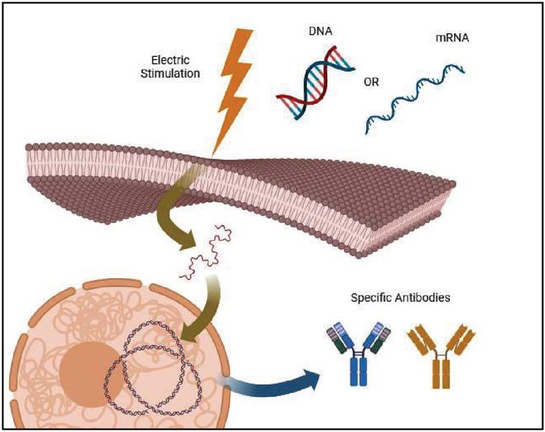
Figure 1 Mechanism of enhancement of the immune response by electroporation.
Innovative Electroporation Applications
In recent years, electroporation has sparked increasing interest and development in the therapeutic field, leading to the emergence of numerous innovative applications. These applications are revolutionizing the treatment of several diseases, improving the effectiveness of existing therapies, and opening up new therapeutic possibilities.
Key areas of innovation include cancer immunotherapy [48], gene therapy [29], cell and tissue therapy [49], antimicrobial therapy, skin and dermatological therapy [50], and neuromodulation [51]. In each of these fields, electroporation has demonstrated significant potential in improving the efficacy of treatments and offering new solutions for difficult-to-treat conditions.
Through electroporation, it has been possible to obtain vaccines that have significantly more marked responses in several clinical trials. Compared to a traditional vaccine approach, the use of DNA plasmids allows for faster vaccine production and lower cost. Furthermore, being stable at room temperature, they can be transported and stored without the need for a cold chain, thus obtaining several advantages from the point of view of emergencies [52], as found during the SARS-CoV-2 pandemic in 2020 [53]. In this particular case, several avenues have been explored, based on mRNA, DNA or inactivated virus vaccines, which involve the use of electroporation to speed up production times [54]. In particular, in the first phase of the epidemic, mRNA-based vaccines were produced, which required a cold chain for the storage and transport phase. For this reason, studies continued, which led to the creation of a plasmid DNA-based vaccine administered through electroporation, with the aim of targeting the Spike protein receptor domain [55]. Through this technique it was possible to observe strong immune and protective responses in animal models, subsequently also being safe and immunogenic for human use [56]. In this way, the aforementioned vaccines were cheaper to produce and facilitated distribution in the various countries where both storage and transport were complex. To make electroporation as effective as possible, a mathematical model was developed that allowed the identification of the main parameters of electric competition. These models have helped to speed up the vaccine production process [57].
Moreover, the utilization of electroporation has enabled a deeper understanding of innovative strategies in cancer therapy, thereby paving the way for the development of a new therapeutic modality that can be employed in vivo. It allows for the introduction of therapeutic genes into cancer cells with the objective of repairing genetic mutations or inducing programmed cell death, as well as triggering specific immune responses [58]. This therapy has undergone significant advancement in the treatment of pancreatic cancer, where the sole traditional curative approach is surgical resection, which is only effective in the early stages of the disease. As documented in the research conducted by Gajewska-Naryniecka et al., the administration of high-voltage pulses can destabilize the cell membrane of diseased cells, ultimately leading to their death [59]. In addition to the pancreas, electroporation can also be employed in the treatment of cancers in other organs, including the prostate. The incidence of prostate cancer has increased significantly in recent years, prompting the development of new, less invasive treatment techniques. As demonstrated by Wang et al., radiofrequency stimulation can induce cancer cell death while selectively sparing the cardiovascular and nervous systems [60].
In addition to the previously referenced works, several studies published in academic literature detail in vivo therapies utilizing irreversible electroporation, with the most contemporary studies outlined in Table 4. This table shows the target organs for therapy, with an emphasis on therapeutic aspects and the applicable treatment periods.
Table 4 Clinical studies that use electroporation as an intervention therapy
| Organ | Therapeutic Aspect | Literature |
| Pancreas | Irreversible electroporation should only be used as an intervention when the patient is presented with locally advanced metastatic disease or is in the initial stages of cancer. In patients with advanced cancer, this treatment is only likely to provide symptom relief. It is important to note that the duration of response to treatment may vary considerably between patients. | [61–63] |
| Prostate | Therapy can be performed when the metastasis is in the initial, localized stage, affecting one or both lobes of the prostate. The treatment has a favorable side-effect profile. | [64, 65] |
| Liver | The treatment may be applied in cases of unresectable liver tumors that do not respond to other clinical treatments or are in the early stages of development. It has been demonstrated that this organ is the most suitable for this treatment. | [66–69] |
| Lungs | The efficacy of the treatment is contingent upon the tumor being unresectable or in an early stage of development. Once more, the procedure is generally well tolerated by the patient, but its efficacy is contingent upon the patient’s clinical status. | [70–72] |
| Breast | The treatment is only applicable in specific cases, namely those where the tumor is diagnosed at an early stage or where surgical intervention is not feasible. It should be noted, however, that this clinical procedure is not applicable to all patients, as there are still contraindications that require further study. | [73–76] |
However, it should be noted that other factors related to the patient’s psycho-physical state of health also influence the efficacy and success of irreversible electroporation therapy.
Challenges in Electroporation: Keeping the Right Local Temperature
The application of high-intensity electrical pulses induces an increase in the temperature of the surrounding biological tissues. The temperature rise is primarily due to the electrical resistance of the tissue to the flow of electric current. When a pulsed electric field is applied, water molecules within the tissue accelerate their movement as they interact with the oscillating electric field. This motion generates friction with neighboring molecules, converting electrical energy into thermal energy through power dissipation processes [77].
The increased temperature can significantly impact the electroporation process and treatment outcomes. Specifically, a moderate temperature rise can enhance cell membrane permeabilization by inducing a lipid phase transition [78]. At higher temperatures, the lipid bilayer of the membrane becomes more fluid, increasing its susceptibility to pore formation and facilitating the entry of molecules. This effect may improve the efficacy of drug or therapeutic agent delivery to target cells [79].
Controlling the temperature rise during electroporation is crucial, as excessively high temperatures can lead to irreversible damage to cells and tissues. Elevated heat levels can cause protein denaturation, rupture of cell membranes, and necrosis in surrounding tissues, thereby compromising both the efficacy and safety of the treatment [80]. The major problems due to the thermal effect and that develop during the treatment are shown in Table 5.
Table 5 Thermal problems of electroporation treatment
| Thermal Effect | Description |
| Overheating of tissues | Application of high-intensity electrical impulses can cause a significant increase in local temperature in surrounding tissues. This overheating can damage cells and tissues, compromising the effectiveness of electroporate treatment and potentially causing undesirable side effects [81]. |
| Protein denaturation | Excessively high temperatures can lead to denaturation of proteins present in tissues. Protein denaturation can alter the structure and function of proteins, negatively affecting biological processes and compromising the health of treated cells [79, 81]. |
| Necrosis of surrounding tissues | Overheating can lead to necrosis, meaning the death of cells in nearby tissue. This is like how intense heat can scorch the edges of a piece of fabric, leaving it weakened or damaged beyond repair. In electroporation treatments, if the surrounding tissue overheats, it can suffer permanent damage, reducing the overall effectiveness of the procedure by damaging areas beyond the intended target [82]. |
| Changes in clinical treatment efficacy | Variations in temperature during the electroporation process can significantly impact the effectiveness of therapeutic agent or molecule delivery within target cells. If temperatures are too high or too low, the transport of these agents across the cell membrane may be hindered, thereby reducing the overall effectiveness of the treatment [83]. |
| Non-specific effects on the cell | High temperatures can induce non-specific effects on treated cells, including alterations to the cell membrane and loss of its structural integrity. These effects can negatively impact cell viability and function, thereby compromising the success of electroporate treatment [79]. |
To mitigate the adverse effects of temperature increases during electroporation, various thermal management techniques are employed [80]. One effective approach is the use of refrigerated electrodes, which dissipate the heat generated during the treatment and help keep the temperature of the treated tissues within safe limits. Another strategy involves cooling the fluid surrounding the tissues to maintain consistent temperature control throughout the electroporation process. Additionally, continuous temperature monitoring during treatment allows for the prompt detection of any unwanted increase, enabling adjustments to the electrical impulse parameters to ensure optimal temperature control and minimize potential damage to surrounding tissues [82–84].
To mitigate the challenges associated with temperature elevation during electroporation, numerous advanced protocols and devices have been developed in recent years [85]. These incorporate the integration of cooling technologies, nanotechnology, computational simulation, and real-time monitoring, elements that have contributed to the advancement of the field of electroporation, enhancing treatment safety and precision [78, 86]. Table 5 presents the principal state-of-the-art protocols, characterized by reduced heat generation and increased clinical efficacy [87]. However, as this technology remains in the developmental stage, certain clinical limitations and operational challenges persist, which are also elucidated in Table 6.
Table 6 Local temperature elevation management methods with clinical cases
| Electroporation | ||
| Type | Description | Clinical Aspect |
| Low frequency pulsed | The low-frequency pulses allow for greater heat dissipation between pulses, thus reducing the risk of overheating. | This method is especially beneficial for treating thermally sensitive organs like the liver and pancreas, where slight temperature rises can reduce therapeutic efficacy or harm nearby tissues [88, 89]. |
| Selective cooling through of nanotechnology | Nanoparticles facilitate heat dissipation from target cells. Researchers are studying gold and silver metal nanoparticles, which exhibit superior thermal conduction compared to biological tissues. | This is emerging as a clinically beneficial strategy, particularly in cancer therapies, where thermal precision is critical to protect surrounding healthy tissues [90, 91]. |
| Pulse reduction in microseconds and nanoseconds | This is another solution to minimize the increase in temperature. Very short pulses, in the order of microseconds or nanoseconds, decrease the amount of energy dissipated as heat. | This has been tested in both animal and in-vitro models for the treatment of tumors, reducing thermal damage and improving tolerability [92, 93]. |
| Cryogenic cooling | This is an experimental technique that aims to limit heat gain through local cooling of the treated area before and after the electrical pulse. | This is used in tumors ablation treatments, where strict thermal control is essential to protect surrounding tissues [62, 94]. |
Analytical Study of Cell Geometry
The study of cell geometry has important implications in several areas of biological and medical research. For instance, understanding the relationship between cell shape and biological functions can provide valuable insights into normal physiology and pathologies associated with alterations in cell shape, such as cancer and degenerative diseases [95]. Indeed, in diseased tissues, changes in the ECM composition and architecture influence the mechanical characteristics and cell geometry [96].
Cell geometry analysis can be used to monitor the efficacy of therapeutic treatments and to develop new strategies for the diagnosis and therapy of diseases [97]. Ultimately, the analytical study of cell geometry represents a powerful approach to explore the complexity and diversity of the cellular world, opening up new perspectives for biomedical research [98].
The analytical study of cell geometry requires a multidisciplinary approach that integrates knowledge from different areas, including biology, mathematics, and computer science. To analyze the geometric characteristics of cells accurately, it is crucial to understand the vast heterogeneity present among different cell types, as well as the variations that can occur in cell size, shape, and structure in response to different biological stimuli or photo-physiological conditions [99].
The approach to cell geometry analysis requires the integration of different methodologies and tools from different disciplines, in order to obtain a comprehensive and detailed understanding of the geometric characteristics of cells and their roles in biological processes [100]. Among the most common methods used for the study of cell geometry is the use of light and electron microscopy, which allows cells to be visualized at high resolution. Fluorescence microscopy, in particular, allows the observation of specific cellular components, such as the nucleus, cytoskeletons and organelles, through the use of fluorescent markers [101, 102]. These images are then processed using dedicated image analysis software, which allows parameters such as area, perimeter, diameter, and complexity of cell shape to be measured [103]. Another technique that allows images to be acquired is scanning electron microscopy (SEM). This technology makes it possible to visualize cells in high resolution, acquiring high-resolution images of their structures. Ultimately, light microscopy and SEM are the tools commonly used to examine cell size and morphology [104].
In previous studies, advanced mathematical models and algorithms for quantitative analysis of cell geometry have been developed. These mathematical models may describe the shape of cells in an accurate and reproducible way, providing detailed information about their structure and geometric properties [105]. For example, some algorithms can identify and quantify cell protrusions, such as pseudopodia and phyllopod, which play a crucial role in biological processes, such as cell migration and interaction with the surrounding environment [106].
In particular, the use of specialized software makes it possible to extract relevant geometric parameters directly from microscopic images. These parameters include area, perimeter, eccentricity, and other parameters that provide quantitative information about the shape and size of the cells [107]. These tools allow scientists to conduct in-depth analyses of cell geometry, revealing important details about cell structure and dynamics in various biological contexts. In parallel, mathematical, and computational analysis plays a crucial role in the study of cell geometry [108]. Mathematical models and computational algorithms are developed to describe and simulate the geometric behavior of cells in response to various conditions [109]. These models can help predict and understand the emergent geometric characteristics of cells based on factors such as population density, the surrounding microenvironment, and the mechanical properties of the cells themselves [110].
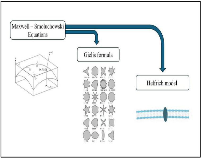
Figure 2 Mathematical model of the cell membrane.
Mathematical Models of the Cell Membrane
Many mathematical models use simplified geometry to describe the biological membrane, considering the membrane as an average non-dispersive structure (Figure 2) [111]. Furthermore, to simplify the model, the thickness of the cell membrane is not taken into consideration. Among the different electroporation methods for simple geometries, a dynamic model concerning pore radii was implemented [112].
In the context of examining the permeability of cell membranes, the three-dimensional quasistatic formulation of Maxwell’s equations, which encompass a set of fundamental partial differential equations in electromagnetism, is applied [16]. By integrating these equations with the nonlinear Smoluchowski equation, which illustrates the dynamics of pores, it is possible to analyze in depth the process of pore formation and evolution within the cell membrane [113].
The laws of dielectric dispersion, which elucidate the behavior of materials under alternating electric fields, have been specifically formulated and incorporated into the numerical code utilized in this study. This incorporation enables the consideration of dispersion effects within the model, thereby enhancing its accuracy and facilitating improved adaptation to the cellular context [114].
To address the complexity of pore dynamics and membrane conductivity, a non-local mathematical model is adopted. In this context, pore density is dependent on trans-membrane tension in a non-direct way, i.e. pore density is not only determined by the applied tension, but also by indirect factors influencing the membrane system [20]. This approach allows for more accurate modelling of the spatial distribution of membrane conductivity, considering the non-local interactions that influence the formation and evolution of pores in the cell membrane [115].
The irregular geometry of the cell can be managed, for example, by the Gielis formula. This formula, based on the concept of the Super formula, represents a mathematical expression that allows the generation of complex and irregular geometric shapes [116]. This formula, developed by mathematician Johan Gielis, incorporates geometric parameters and trigonometric functions to describe a wide range of shapes, which go beyond classic geometric figures such as circles, squares, or ellipses [117].
In the context of cell geometry, the application of the Gielis formula makes it possible to accurately model the complex morphology of cells, considering their structural and morphological variations in different biological contexts. This mathematical model can be used to accurately analyze and represent the shape of cells in biological tissues, cell cultures, or other experimental environments [118].
Using the Gielis formula allows scientists to obtain a detailed mathematical representation of the geometric characteristics of cells, providing useful information for studying biological processes such as morphogenesis, cell migration, and cell interaction with the surrounding environment. In addition, it allows us to deal with the complexity of cell geometry in a rigorous and accurate way, thus contributing to the understanding of biological structures at a mathematical and computational level [119].
Finally, statistical analysis is often employed to assess geometric variability between different cell populations or experimental conditions. Advanced statistical techniques are used to compare and analyze geometric data from different sources, allowing any correlations or significant differences in cell geometry to be identified [120].
Theoretical frameworks grounded in mathematical models are essential for investigating the composition and traits of cell membranes. These models offer a precise and comprehensive depiction of the intricate molecular and subcellular interactions that characterize the cell membrane. By employing differential equations and advanced computational simulations, important phenomena, such as lipid fluidity, selective permeability, and molecular transport across the membrane, can be accurately modelled. This method enables a thorough analysis of the molecular mechanisms that govern the activities of the cell membrane, providing valuable insights into its structure and functionality. There are several reasons for developing mathematical models of cell membranes. First, the models enable the synthesis and organization of current knowledge about the structure and functioning of membranes, allowing for a deeper understanding of such intricate biological systems. Second, the models allow for the prediction of membrane behavior in response to changes in environmental conditions or experimental manipulations, enabling quantitative predictions to be made and scientific hypotheses to be tested. Finally, the development of mathematical models can guide the design of future experiments and the identification of new areas of research, thereby helping to advance our understanding of cell biology.
The ability to predict the behavior of cell membranes in response to external stimuli or changes in physiological conditions is one of the main advantages of mathematical models [121]. These models allow a wide range of scenarios to be explored virtually, anticipating membrane responses to changes in ion concentration, transmembrane electrical potentials, or exposure to chemical or physical agents [122]. Through the analysis of simulations, it is possible to identify the molecular mechanisms underlying membrane responses, better understanding the transport processes and protein–lipid interactions that regulate cell function [123]. In addition, mathematical models allow the exploration of the role of cell membranes in more biological contexts, such as in cell–cell interactions or cell communication. The ability to model membrane dynamics within complex systems provides valuable insights into the regulation of biological processes and can guide the development of targeted therapies or diagnostic strategies [124].
This includes simulating certain phenomena, such as osmosis, ion balance, and various ion channel activities. In addition, the mathematical models make it possible to accurately represent the complex interactions that take place in and around cell membranes [123]. These models take into account a wide range of factors, including the lipid composition of the membrane, the presence and distribution of transmembrane proteins, ion concentration gradients, and electrical potentials across the membrane [125]. Through mathematical equations, we can describe the selective permeability of the membrane, the transport of molecules through it, and the molecular interactions that occur on its surface [126].
Cell membranes, consisting of a phospholipid bilayer, represent a complex structure characterized by a hydrophilic polar head and a hydrophobic lipid tail. Their thin size, typically between 5 and 100 nanometers, makes them ideal for being modelled effectively through two-dimensional approaches [123, 127]. This approximation is supported by the observation of the radii of curvature of the cell surfaces, which are much greater than the thickness of the membrane, suggesting that the deformation occurs mainly on the outer surface [128].
Since cells can take heterogeneous shapes, the first step in the study of the membrane zone involves the analysis of cell geometry and flexural factors [129]. To this end, several advanced theoretical models have been developed over the years, including the “spontaneous curvature” model, which uses the equation of the bending energy of the vesicle and the Helfrich model. In particular, the latter model considers the membrane as a two-dimensional elastic surface subject to deformation. The first step in deriving geometry is to determine the total free energy of a deformed membrane. This energy can be expressed through Equation (1).
| (1) |
The parameters in the equation represent:
• represents the flexural modulus of the membrane.
• and represent the main curvatures of the membrane.
• represents spontaneous curvature. This parameter is very important, as it will take into account how the membrane is structured, such as its thickness and the number of layers that compose it.
• represents Gauss’s modulus, or torsion constant. If the deformation turns out to be planar, it is overlooked.
• as a stress profile. For simplify the models, this parameter was considered trans-monolayer.
This model provides a mathematical description of the relationship between membrane curvature and associated energy, thus offering a theoretical framework for understanding the different flexural properties of cell membranes.
To define the theoretical membrane shape, it is necessary to derive the minimum of the function , which depends on both an area and a volume bound to the cell membrane. To solve this problem, we introduce the Lagrange multipliers , introducing relation (2).
| (2) |
The parameter indicates the change in the function , and represents the difference in pressure between the outside and inside of the membrane.
Once the above equations have been defined, it will be necessary to introduce relationships, based on the type of reference system, which will allow the geometry of the membrane to be defined. This will lead to the formulation of a system of differential equations, which allows the shape of the cell membrane to be determined. However, because the problem has more unknowns than equations, you will need to set appropriate boundary and edge conditions for its solution [130].
Examples of the use of Helfrich’s model and the equation of membrane liberated energy can be found in several studies on the biomechanics of cell membranes and the morphology of lipid vesicles [131]. For example, a study conducted by Seifert and Lipoweky applied Helfrich’s model to analyze the deformation of lipid vesicles subject to adhesion and hydrodynamic flow. Using this model, it was investigated how the interaction between vesicles and solid surfaces influenced the shape and dynamics of lipid vesicles [132].
A recent study conducted by Bernard and colleagues presents a mathematical model that focuses on spherical red blood cells, with the intent of deepening the understanding of spherocytes and spherocytosis. The paper provides a detailed classification of spherical solutions based on Helfrich’s model, and includes an in-depth analysis of membrane stiffness and spherocyte stability [133].
The work conducted by Basesu et al. introduced an advanced mathematical model to examine the finite deformation of cell membranes and to better understand their local mechanical response. The proposed model is based on the use of an isothermal function of free energy, derived from Helfrich functions, per unit mass. This function is formulated based on the scalar invariants of surface deformation and curvature. This approach allows an accurate representation of the mechanical response of cell membranes, taking into account their surface structure and the forces acting on them [131].
Subsequently, further mathematical models describing the mechanical properties and dynamics of cell membranes were developed. For instance, the Canham–Helfrich model extends Helfrich’s formalism by introducing an additional term () inherent in mean surface tension into the formulation. This model was initially developed to explain the biconcave shape of the red blood cell and then applied to other types of cells. The Canham–Helfrich equation is reported, updated with the supplementary term , in (3).
| (3) |
This term is proportional to the difference between the instantaneous mean curvature and the reference curvature. In particular, the mean curvature is defined starting from the main curvatures (first term), in order to describe the large-scale geometry of the membrane, such as the change in shape due to the presence of localized curvatures [134].
Through the above relationship, it is possible to derive a free energy density that allows us to define a spontaneous mean curvature that includes information regarding the preferred natural form of the lipid bilayer.
Some studies use the Canham–Helfrich relationship to analyze the morphology and mechanical characteristics of cell membranes in order to understand phenomena such as vesicle formation, cell morphology, and membrane fusion and fission processes. For instance, the work by Seguin and Fried focuses on the derivation of the Canham–Helfrich relation to explain the origin of spontaneous curvature, attributing it to the axial asymmetry between the sheets constituting the lipid bilayer [135]. In another research the distribution of the phase field was analyzed to manage pearling instabilities, resulting from the anchoring of amphiphilic polymers on a lipid vesicle. The primary objective of the research was to identify and analyze the morphological instabilities observed through examples, to obtain a deeper understanding of the underlying mechanisms that regulate their appearance. It was shown that by integrating the experimental data with the application of a polymer on a global scale, using the Canham–Helfrich relation, at a concentration of polymer anchors, the homogeneous pearl configuration is less energetically favorable than an inhomogeneous configuration [136].
However, although the developed models are easy to interpret, they present significant limitations at the computational level, resulting in difficulties in accurately solving simulations. In particular, the algorithmic complexity and the high demand for computational resources can compromise numerical accuracy and stability, especially when dealing with phenomena that require high spatial or temporal resolution.
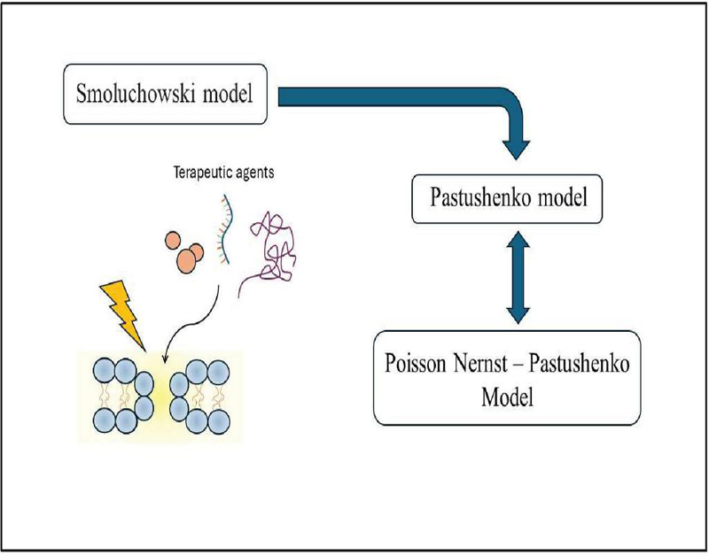
Figure 3 Mathematical models for electroporation.
Mathematical Models for Electroporation
Electroporation has been the subject of numerous experimental studies and has found a wide range of practical applications in areas such as gene therapy, drug delivery, and cell manipulation [137]. However, despite significant progress in the understanding and practical application of electroporation, its theoretical aspect remains a challenge [138].
There are several reasons why the theoretical aspect of electroporation is complex and presents significant challenges:
• Complexity of physical interactions: Electroporation involves complex interactions between electric fields, cell membranes, and intracellular and extracellular fluids. These interactions can be highly nonlinear and dependent on numerous factors, such as the strength of the electric field, the duration of the electrical impulses, the chemical composition of the cell membrane, and more [138, 139].
• Variations between biological systems: The properties of cell membranes and the response of cells to electroporation can vary greatly between different cell types, tissues, and biological species. This variation complicates the formulation of theoretical models that can be applied universally and requires a more detailed and specific analysis for each biological system [140].
• Limitations in experimental data: Although electroporation has been extensively studied experimentally, there are still gaps in the available data and detailed information about the exact dynamics of the phenomenon. These limitations can complicate the development and validation of accurate theoretical models [8].
Understanding the exact mechanisms by which cell membranes undergo and react to electroporation is still the subject of active research.
The first mathematical model developed to simulate the behavior of the cell membrane is based on the use of the Smoluchowski equation (Figure 3, Equation (4)) [141]. This differential equation makes it possible to model the kinetics of chemical reactions and the transport of particles in the environment surrounding the cell membrane, as well as to simulate the responses of the membrane itself [142]. In particular, the differential equation makes it possible to plot the trend over time t of the probability density that a particle is in a specific position and time instant.
| (4) |
In Equation (4), is the probability density of finding a particle at position and at time is the diffusion coefficient, which has the following form where is Boltzmann’s constant, is the temperature, the kinematic viscosity of the fluid medium, and is the radius of the particle, and represents the field of forces acting on the particle [143, 144].
However, although it is a useful model for describing the diffusion of particles in a field of external forces, it may encounter some limitations when applied to electroporation. First, the equation assumes that the particles behave like classical particles, ignoring any quantum or correlation effects between particles that might contain their motion [145, 146]. This limitation can be significant in small systems, such as nanoparticles or nanomaterials involved in electroporation. To solve these challenges, analytical and numerical models have been developed that allow electroporation and its effects on different scenarios to be simulated [16]. These models allow for both qualitative and quantitative characterization of the onset and dynamics of cell pores, allowing you to examine how they vary in response to factors such as electric field strength and the duration of electrical impulses [147]. Another challenge concerns the interaction of particles with the electric field applied during electroporation. Although the Smoluchowski equation includes a term for external forces such as the electric field, the complexity and strength of the electric field during electroporation can cause significant changes in the cell membrane and the behavior of charged particles [148]. These effects cannot be adequately modelled by Smoluchowski’s approach, which assumes a linear dependence between the electric field and the resulting force on the particles. In addition, during electroporation, the electric field can vary in time and space, especially with the use of short, intense electrical pulses [149]. The Smoluchowski equation may not be able to accurately capture these dynamical variations in the electric field, which can significantly vary the behavior of particles and their interaction with the cell membrane [150]. Phenomena such as cell membrane polarization, which occur when the electric field induces changes in charge distribution on the membrane, can also interfere with the transfer of particles across the membrane during electroporation. These effects cannot be adequately modelled by the Smoluchowski equation, which does not take into account the specific interactions between particles and the cell membrane [151].
Subsequently, more sophisticated models were introduced, such as the Pastushenko model, which turns out to be closely related to electroporation [152]. The above relation is a partial differential equation that describes the transport of charged particles, such as ions or molecules, across a cell membrane subjected to an external electric field [77]. Equation (5) provides a mathematical description of the balance between the diffusive flux of particles, governed by the concentration gradient, and the convective flux caused by the effect of the electric field charge.
| (5) |
In Equation (5), parameter c indicates the concentration of the ionic or molecular species of interest, q indicates the charge of the species, and indicates the applied electric potential.
This equation represents the balance between the diffusive flux of the particles, governed by the concentration gradient (first term), and the convective flux due to the presence of the electric field (second term). The first term indicates the net flux of particles due to their tendency to move from the region of greater to that of lower concentration. The second term represents the flux because of the electric field on charged particles, which pushes them to cross the cell membrane [153].
In the context of electroporation, the Pastushenko equation can be used to model the transport of ions, molecules, or nanoparticles across the cell membrane during the application of electrical impulses. The diffusive term of the equation considers the random motion of particles due to their tendency to move from regions of higher to lower concentration. This is particularly relevant during electroporation, as the application of electrical pulses can cause a temporary increase in membrane permeability, allowing particles to pass through temporarily formed pores [154].
On the other hand, the convective term of Pastushenko’s equation represents the particle flux induced by the interaction with the electric field applied during electroporation. When the particles are charged, the electric field exerts a force on them, causing them to cross the cell membrane. This convective effect is crucial to facilitate the transport of particles across the membrane during electroporation and can be effectively described by the Pastushenko equation [155].
However, the practical implementation of this model has some limitations and challenges that can affect its validity and accuracy in fully describing the phenomenon [25]. One of the main challenges is the simplifications inherent in the model. Pastushenko’s equation assumes a uniform and isotropic cell membrane, neglecting heterogeneity or complex structures present in the membrane that could significantly influence particle behavior [156]. This can lead to an inaccurate representation of transport across the membrane, especially under non-uniform conditions or in the presence of specific regions of the membrane that are more permeable or resistive [157].
Another important limitation concerns the applicability of the Nernst–Planck equation, on which the Pastushenko equation is based, to describe the diffusive flow of particles. The Nernst–Planck equation is only valid under certain conditions, such as low concentration gradients and low electric field values [158]. In situations where high concentration gradients and intense electric fields are present, the Nernst–Planck equation may not be accurate enough, and more complex models, such as the Poisson–Nernst–Planck equation, which considers the effects of electric force on ion transport, may be required [159]. In addition, the Pastushenko equation may not take into account complex phenomena that can occur during electroporation, such as the formation and dynamics of pores in the membrane, interaction with membrane proteins, or cell membrane polarization [160]. These factors can significantly affect particle transport and may require more detailed models to be adequately represented [161].
Another aspect related to the problems on the development of theoretical models for electroporation is the complexity of the multiscale interactions involved, which include electric fields, cell membranes and intracellular and extracellular fluids [162]. These interactions require a rigorous mathematical approach to be accurately described. In addition, the presence of heterogeneity in biological tissues and variations in the properties of cell membranes add further challenges to theoretical modelling, as such variations need to be taken into account in models [163, 164]. In addition, the implementation of such models requires significant computational power and long computational times, especially when considering the complexity of the interactions and the need to include multiple variables and boundary conditions [165]. Therefore, an important goal in the development of such models is the search for efficient computational methods that can reduce computational cost and computational time without compromising the accuracy of simulations [166, 167].
Challenges and Conclusions
Electroporation is an innovative therapeutic approach in the biomedical field, with significant applications in cancer treatment and targeted drug delivery. This technique utilizes electrical impulses to increase cell membrane permeability, facilitating the entry of molecules with specific geometric characteristics into target cells. Preclinical and clinical studies indicate that electroporation enhances therapeutic efficacy and reduces systemic side effects, representing a significant advancement in cancer treatment strategies. This method delivers chemotherapeutic agents or other therapeutic molecules directly into tumor cells, enhancing treatment effectiveness while minimizing systemic side effects compared with conventional therapies. Furthermore, it enhances drug transport across cell membranes, thus enabling more targeted and efficacious treatments. This is particularly advantageous for conditions necessitating high specificity in therapeutic action.
Clinical application of electroporation faces technical and biological challenges. Electrical impulses can increase tissue temperature, potentially causing non-specific cell damage and compromising treatment safety, particularly in proximity to vital organs. The requisite current intensity to achieve the desired effect varies by tissue type and treatment conditions, complicating protocol standardization. High current intensity can also elicit side effects such as pain and involuntary muscle contractions, affecting patient acceptability and tolerability.
Developing mathematical models to describe cell membrane behavior during electroporation is crucial. These models enhance understanding of biomedical mechanisms, simulate electrical impulse effects on cells, and predict optimal pore-opening conditions. Integrating experimental data with theoretical analyses refined treatment parameters, reducing thermal damage risks and improving therapeutic efficacy. Customizable models can account for individual biological variations, such as cell membrane composition and target tissue characteristics, thereby improving targeted therapeutic approaches and clinical outcomes. However, creating and validating models present challenges due to computational and biological factors. A comprehensive understanding of cell membrane biophysics and electrical interactions, along with advanced mathematical and computational skills, is required. Acquiring high-quality experimental data for model validation turns out to be challenging, while the biological variability of models complicates the production of effective for the equivalent clinical contexts.
Recent advancements in electroporation include novel vectors for drug and nucleic acid transport, enhancing delivery efficacy and selectivity while reducing diffusion to healthy tissues. Innovative technologies have facilitated bespoke clinical applications in drug transport and cancer therapy. Clinical studies demonstrate efficacy in treating tumors, infections, and genetic diseases, thus revealing new therapeutic possibilities. The development of targeted and customized protocols for therapeutic treatments is essential, as current protocols may not be suitable for all patients, necessitating tailored approaches for optimal treatment outcomes.
In summary, electroporation is a promising therapeutic approach with significant potential. However, additional research is necessary to reduce its potential negative consequences. The ongoing optimization of treatment parameters and the development of more precise and manageable electroporation systems, aided by advanced mathematical models, are critical to realizing the therapeutic potential of this technology while ensuring the highest standards of patient safety and well-being. The incorporation of new knowledge and technologies could thus facilitate the definitive establishment of electroporation as a leading therapeutic tool in contemporary medicine.
References
[1] S. J. Singer e G. L. Nicolson, «The Fluid Mosaic Model of the Structure of Cell Membranes», Science, vol. 175, fasc. 4023, pp. 720–731, feb. 1972, doi: 10.1126/science.175.4023.720.
[2] K. Simons e E. Ikonen, «Functional rafts in cell membranes», Nature, vol. 387, fasc. 6633, pp. 569–572, giu. 1997, doi: 10.1038/42408.
[3] Lingwood, D., and Simons, K. (2010). Lipid rafts as a membrane-organizing science, 327(5961), 46–50.
[4] W. A. Catterall, «From Ionic Currents to Molecular Mechanisms: The Structure and Function of Voltage-Gated Sodium Channels», Neuron, vol. 26, fasc. 1, pp. 13–25, apr. 2000, doi: 10.1016/S0896-6273(00)81133-2.
[5] Staub, O., and Rotin, D. (2006). Role of ubiquitylation in cellular membrane transport. Physiological reviews, 86(2), 669–707.
[6] W. A. Catterall, «Structure and Regulation of Voltage-Gated Ca2+ Channels», Annual Review of Cell and Developmental Biology, vol. 16, fasc. Volume 16, 2000, pp. 521–555, nov. 2000, doi: 10.1146/annurev.cellbio.16.1.521.
[7] S. Tan, T. Wu, D. Zhang, e Z. Zhang, «Cell or Cell Membrane-Based Drug Delivery Systems», Theranostics, vol. 5, fasc. 8, p. 863, 2015, doi: 10.7150/thno.11852.
[8] T. Batista Napotnik, T. Polajžer, e D. Miklavèiè, «Cell death due to electroporation – A review», Bioelectrochemistry, vol. 141, p. 107871, ott. 2021, doi: 10.1016/j.bioelechem.2021.107871.
[9] T. Kotnik, P. Kramar, G. Pucihar, D. Miklavcic, e M. Tarek, «Cell membrane electroporation- Part 1: The phenomenon», IEEE Electrical Insulation Magazine, vol. 28, fasc. 5, pp. 14–23, set. 2012, doi: 10.1109/MEI.2012.6268438.
[10] M. A. Chiapperino, P. Bia, C. M. Lamacchia, e L. Mescia, «Electroporation Modelling of Irregular Nucleated Cells Including Pore Radius Dynamics», Electronics, vol. 8, fasc. 12, Art. fasc. 12, dic. 2019, doi: 10.3390/electronics8121477.
[11] C. Zhou, Z. Yan, e K. Liu, «Response characteristics and optimization of electroporation: simulation based on finite element method», Electromagnetic Biology and Medicine, vol. 40, fasc. 3, pp. 321–337, lug. 2021, doi: 10.1080/15368378.2021.1951484.
[12] R. P. Joshi e K. H. Schoenbach, «Electroporation dynamics in biological cells subjected to ultrafast electrical pulses: A numerical simulation study», Phys. Rev. E, vol. 62, fasc. 1, pp. 1025–1033, lug. 2000, doi: 10.1103/PhysRevE.62.1025.
[13] E. Langhoff e J. Ladefoged, «Sodium activity, sodium concentration, and osmolality in plasma in acute and chronic renal failure», Clin Chem, vol. 31, fasc. 11, pp. 1811–1814, nov. 1985.
[14] M. Zacchia, M. L. Abategiovanni, S. Stratigis, e G. Capasso, «Potassium: From Physiology to Clinical Implications», Kidney Diseases, vol. 2, fasc. 2, p. 72, mag. 2016, doi: 10.1159/000446268.
[15] T. Kotnik, L. Rems, M. Tarek, e D. Miklavèiè, «Membrane Electroporation and Electropermeabilization: Mechanisms and Models», Annual Review of Biophysics, vol. 48, fasc. Volume 48, 2019, pp. 63–91, mag. 2019, doi: 10.1146/annurev-biophys-052118-115451.
[16] Goldberg, E., Suárez, C., Alfonso, M., Marchese, J., Soba, A., and Marshall, G. (2018). Cell membrane electroporation modeling: A multiphysics Bioelectrochemistry, 124, 28–39.
[17] Napotnik, T. B., and Miklavèiè, D. (2018). In vitro electroporation detection methods – An overview. Bioelectrochemistry, 120, 166–182.
[18] Liu, J., Jiang, J., He, M., Chen, J., Huang, S., Liu, Z., … and Wang, J. (2023). Nanopore Electroporation Device for DNA Transfection into Various Spreading and Nonadherent Cell Types. ACS Applied Materials & Interfaces, 15(43), 50015–50033.
[19] Zeller, R. W. (2018). Electroporation in ascidians: history, theory and Transgenic ascidians, 37–48.
[20] G. Pucihar, T. Kotnik, e D. Miklavèiè, «Measuring the Induced Membrane Voltage with Di-8-ANEPPS», JoVE (Journal of Visualized Experiments), fasc. 33, p. e1659, nov. 2009, doi: 10.3791/1659.
[21] Gehl, J. J. A. P. S. (2003). Electroporation: theory and methods, perspectives for drug delivery, gene therapy and research. Acta Physiologica Scandinavica, 177(4), 437–447.
[22] S. Skorupska, I. Grabowska-Jadach, A. Dybko, e Z. Brzozka, «Studies on electroporation and electrochemotherapy of adherent cells monolayer using electrode modules of specific geometry», Sensors and Actuators B: Chemical, vol. 351, p. 130889, gen. 2022, doi: 10.1016/j.snb.2021.130889.
[23] Evangelopoulos, M., Yazdi, I. K., Acciardo, S., Palomba, R., Giordano, F., Pasto, A., … and Tasciotti, E. (2020). Biomimetic cellular vectors for enhancing drug delivery to the lungs. Scientific Reports, 10(1), 172.
[24] McBride, J. L., Boudreau, R. L., Harper, S. Q., Staber, P. D., Monteys, A. M., Martins, I., … and Davidson, B. L. (2008). Artificial miRNAs mitigate shRNA-mediated toxicity in the brain: implications for the therapeutic development of Proceedings of the National Academy of 105(15), 5868–5873.
[25] Neumann, E., SchaeferRidder, M., Wang, Y., and Hofschneider, P. (1982). Gene transfer into mouse lyoma cells by electroporation in high electric fields. The EMBO journal, 1(7), 841–845.
[26] M. Breton e L. M. Mir, «Microsecond and nanosecond electric pulses in cancer treatments», Bioelectromagnetics, vol. 33, fasc. 2, pp. 106–123, 2012, doi: 10.1002/bem.20692.
[27] Sersa, G., Kranjc, S., Scancar, J., Krzan, M., and Cemazar, M. (2010). Electrochemotherapy of mouse sarcoma tumors using electric pulse trains with repetition frequencies of 1 Hz and 5 kHz. The Journal of membrane biology, 236, 155–162.
[28] Zhang, Z., Huang, G., Li, Y., Chen, X., Yao, Y., Ren, S., … and An, C. (2022). Electrically conductive inorganic membranes: A review on principles, characteristics and applications. Chemical Engineering Journal, 427, 131987.
[29] F. Liu, R. Su, X. Jiang, S. Wang, W. Mu, e L. Chang, «Advanced micro/nano-electroporation for gene therapy: recent advances and future outlook», Nanoscale, vol. 16, fasc. 22, pp. 10500–10521, giu. 2024, doi: 10.1039/D4NR01408A.
[30] K. Balantiè, D. Miklavèiè, I. Križaj, e P. Kramar, «The Good and the Bad of Cell Membrane Electroporation», ACSi, vol. 68, fasc. 4, pp. 753–764, dic. 2021, doi: 10.17344/acsi.2021.7198.
[31] Spugnini, E. P., Condello, M., Crispi, S., and Baldi, A. (2024). Electroporation in Translational Medicine: From Veterinary Experience to Human Oncology. Cancers, Cancers, 16(5), 1067.
[32] Bo, W., Silkunas, M., Mangalanathan, U., Novickij, V., Casciola, M., Semenov, I., … and Pakhomov, A. G. (2020). Probing Nanoelectroporation and Resealing of the Cell Membrane by the Entry of Ca and Ba Ions. International Journal of Molecular Sciences, 21(9), 3386.
[33] Vorobiev, E., Lebovka, N., Vorobiev, E., and Lebovka, N. (2020). Fundamentals of electroporation, theory and mathematical models for simulation of PEE processing. Processing of Foods and Biomass Feedstocks by Pulsed Electric Energy, 27–49.
[34] S. Chopra, P. Ruzgys, M. Maciulevièius, M. Jakutavièiûtë, e S. Ðatkauskas, «Investigation of Plasmid DNA Delivery and Cell Viability Dynamics for Optimal Cell Electrotransfection In Vitro», Applied Sciences, vol. 10, fasc. 17, Art. fasc. 17, gen. 2020, doi: 10.3390/app10176070.
[35] Y. Cu et al., «Enhanced Delivery and Potency of Self-Amplifying mRNA Vaccines by Electroporation in Situ», Vaccines (Basel), vol. 1, fasc. 3, pp. 367–383, ago. 2013, doi: 10.3390/vaccines1030367.
[36] R. S. Son, K. C. Smith, T. R. Gowrishankar, P. T. Vernier, e J. C. Weaver, «Basic Features of a Cell Electroporation Model: Illustrative Behavior for Two Very Different Pulses», J Membrane Biol, vol. 247, fasc. 12, pp. 1209–1228, dic. 2014, doi: 10.1007/s00232-014-9699-z.
[37] Chen, Y., Moser, M. A., Luo, Y., Zhang, W., and Zhang, B. (2019). Chemical enhancement of irreversible electroporation: A review and future suggestions. Technology in Cancer Research & Treatment, 18, 1533033819874128.
[38] Chang, A. Y., Liu, X., Tian, H., Hua, L., Yang, Z., and Wang, S. (2020). Microfluidic electroporation coupling pulses of nanoseconds and milliseconds to facilitate rapid uptake and enhanced expression of DNA in cell therapy. Scientific reports, 10(1), 6061.
[39] L. Lambricht, A. Lopes, S. Kos, G. Sersa, V. Préat, e G. Vandermeulen, «Clinical potential of electroporation for gene therapy and DNA vaccine delivery», Expert Opinion on Drug Delivery, vol. 13, fasc. 2, pp. 295–310, feb. 2016, doi: 10.1517/17425247.2016.1121990.
[40] K. E. Broderick e L. M. Humeau, «Electroporation-enhanced delivery of nucleic acid vaccines», Expert Review of Vaccines, vol. 14, fasc. 2, pp. 195–204, feb. 2015, doi: 10.1586/14760584.2015.990890.
[41] J. Jiang et al., «Integration of needle-free jet injection with advanced electroporation delivery enhances the magnitude, kinetics, and persistence of engineered DNA vaccine induced immune responses», Vaccine, vol. 37, fasc. 29, pp. 3832–3839, giu. 2019, doi: 10.1016/j.vaccine.2019.05.054.
[42] K. R. Porter e K. Raviprakash, «DNA Vaccine Delivery and Improved Immunogenicity», Current Issues in Molecular Biology, vol. 22, fasc. 1, Art. fasc. 1, apr. 2017, doi: 10.21775/cimb.022.129.
[43] Ledesma-Feliciano, C., Chapman, R., Hooper, J. W., Elma, K., Zehrung, D., Brennan, M. B., and Spiegel, E. K. (2023). Improved DNA vaccine delivery with needle-free injection systems. Vaccines, 11(2), 280.
[44] Lu, B., Lim, J. M., Yu, B., Song, S., Neeli, P., Sobhani, N., … and Chai, D. (2024). The next-generation DNA vaccine platforms and delivery systems: Advances, challenges and prospects. Frontiers in immunology, 15, 1332939.
[45] J. B. Sandbrink e G. D. Koblentz, «Biosecurity risks associated with vaccine platform technologies», Vaccine, vol. 40, fasc. 17, pp. 2514–2523, apr. 2022, doi: 10.1016/j.vaccine.2021.02.023.
[46] D. N. Kisakov, I. M. Belyakov, L. A. Kisakova, V. A. Yakovlev, E. V. Tigeeva, e L. I. Karpenko, «The use of electroporation to deliver DNA-based vaccines», Expert Review of Vaccines, vol. 23, fasc. 1, pp. 102–123, dic. 2024, doi: 10.1080/14760584.2023.2292772.
[47] A. Cvetkoska, E. Pírc, M. Reberšek, R. Magjarević, e D. Míklavèiè, «Towards standardization of electroporation devices and protocols», IEEE Instrumentation & Measurement Magazine, vol. 23, fasc. 2, pp. 74–81, apr. 2020, doi: 10.1109/MIM.2020.9062692.
[48] Pisani, S., Bertino, G., Prina-Mello, A., Locati, L. D., Mauramati, S., Genta, I., … and Benazzo, M. (2022). Electroporation in head-and-neck cancer: an innovative approach with immunotherapy and nanotechnology combination. Cancers, Cancers, 14(21), 5363.
[49] Rakoczy, K., Kisielewska, M., Sêdzik, M., Jonderko, L., Celińska, J., Sauer, N., … and Kulbacka, J. (2022). Electroporation in clinical applications – The potential of gene electrotransfer and electrochemotherapy. Applied Sciences, 12(21), 10821.
[50] R. Upasani, A. Herwadkar, N. Singh, e A. K. Banga, «Innovations and Future Prospects of Dermal Delivery Systems», in Dermal Drug Delivery, CRC Press, 2020.
[51] M. Liu et al., «Recent advances and research progress on microsystems and bioeffects of terahertz neuromodulation», Microsyst Nanoeng, vol. 9, fasc. 1, pp. 1–17, nov. 2023, doi: 10.1038/s41378-023-00612-1.
[52] F. Saade e N. Petrovsky, «Technologies for enhanced efficacy of DNA vaccines», Expert Review of Vaccines, vol. 11, fasc. 2, pp. 189–209, feb. 2012, doi: 10.1586/erv.11.188.
[53] Amanat, F., and Krammer, F. (2020). SARS-CoV-2 vaccines: status Immunity, 52(4), 583–589.
[54] Z. Xu et al., «Harnessing Recent Advances in Synthetic DNA and Electroporation Technologies for Rapid Vaccine Development Against COVID-19 and Other Emerging Infectious Diseases», Front. Med. Technol., vol. 2, ott. 2020, doi: 10.3389/fmedt.2020.571030.
[55] F. D’Alessio et al., «Immunogenicity of COVID-eVax Delivered by Electroporation Is Moderately Impacted by Temperature and Molecular Isoforms», Vaccines, vol. 11, fasc. 3, Art. fasc. 3, mar. 2023, doi: 10.3390/vaccines11030678.
[56] L. Aurisicchio et al., A first-in-human trial on the safety and immunogenicity of COVID-eVax, a cellular response-skewed DNA vaccine against COVID-19, Mol Ther, vol. 31, fasc. 3, pp. 788–800, mar. 2023, doi: 10.1016/j.ymthe.2022.12.017.
[57] A Mathematical Model to Expedite Electroporation Based Vaccine Development for COVID-19, Biointerface Res Appl Chem, vol. 12, fasc. 2, pp. 1951–1961, giu. 2021, doi: 10.33263/BRIAC122.19511961.
[58] C. He, X. Huang, Y. Zhang, X. Lin, e S. Li, T-cell activation and immune memory enhancement induced by irreversible electroporation in pancreatic cancer, Clinical and Translational Medicine, vol. 10, fasc. 2, p. e39, 2020, doi: 10.1002/ctm2.39.
[59] A. Gajewska-Naryniecka, U. Szwedowicz, Z. Łapińska, J. Rudno-Rudzińska, W. Kielan, e J. Kulbacka, Irreversible Electroporation in Pancreatic Cancer – An Evolving Experimental and Clinical Method, International Journal of Molecular Sciences, vol. 24, fasc. 5, Art. fasc. 5, gen. 2023, doi: 10.3390/ijms24054381.
[60] H. Wang et al., «Extended Focal Ablation of Localized Prostate Cancer With High-Frequency Irreversible Electroporation: A Nonrandomized Controlled Trial», JAMA Surgery, vol. 157, fasc. 8, pp. 693–700, ago. 2022, doi: 10.1001/jamasurg.2022.2230.
[61] P. Charalambous et al., «The efficacy and safety of the open approach irreversible electroporation in the treatment of pancreatic cancer: A systematic review», European Journal of Surgical Oncology, 46, fasc. 9, pp. 1565–1572, set. 2020, doi: 10.1016/j.ejso.2020.05.017.
[62] Z. L. Rai, R. Feakins, L. J. Pallett, D. Manas, e B. R. Davidson, Irreversible Electroporation (IRE) in Locally Advanced Pancreatic Cancer: A Review of Current Clinical Outcomes, Mechanism of Action and Opportunities for Synergistic Therapy, Journal of Clinical Medicine, vol. 10, fasc. 8, Art. fasc. 8, gen. 2021, doi: 10.3390/jcm10081609.
[63] A. Gyftopoulos, I. A. Ziogas, A. S. Barbas, e D. Moris, The Synergistic Role of Irreversible Electroporation and Chemotherapy for Locally Advanced Pancreatic Cancer, Front. Oncol., vol. 12, mag. 2022, doi: 10.3389/fonc.2022.843769.
[64] A. Kiełbik, W. Szlasa, J. Saczko, e J. Kulbacka, Electroporation-Based Treatments in Urology, Cancers, vol. 12, fasc. 8, Art. fasc. 8, ago. 2020, doi: 10.3390/cancers12082208.
[65] L. Lunelli, O. Cussenot, e J. J. M. C. H. de la Rosette, «Irreversible Electroporation (IRE) for Prostate Cancer», in Interventional Urology, A. R. Rastinehad, D. N. Siegel, B. J. Wood, e T. McClure, A c. di, Cham: Springer International Publishing, 2021, pp. 241–247. doi: 10.1007/978-3-030-73565-4_16.
[66] Yu, M., and Li, S. (2022). Irreversible electroporation for liver cancer ablation: A meta analysis. European Journal of Surgical Oncology, 48(6), 1321–1330.
[67] Z.-G. Liu, X.-H. Chen, Z.-J. Yu, J. Lv, e Z.-G. Ren, «Recent progress in pulsed electric field ablation for liver cancer», World J Gastroenterol, vol. 26, fasc. 24, pp. 3421–3431, giu. 2020, doi: 10.3748/wjg.v26.i24.3421.
[68] D. Cano, J. J. Lasarte, e I. Vivas, [Irreversible electroporation: present and future in the treatment of hepatocellular carcinoma, An Sist Sanit Navar, vol. 45, fasc. 3, p. e1019, nov. 2022, doi: 10.23938/assn.1019.
[69] J. Yang, A. Eresen, J. Shangguan, Q. Ma, V. Yaghmai, e Z. Zhang, Irreversible electroporation ablation overcomes tumor-associated immunosuppression to improve the efficacy of DC vaccination in a mice model of pancreatic cancer, OncoImmunology, vol. 10, fasc. 1, p. 1875638, gen. 2021, doi: 10.1080/2162402X.2021.1875638.
[70] A. Nikyar e A. Bolhassani, Electroporation: An Effective Method For In Vivo Gene Delivery, Drug Delivery Letters, vol. 12, fasc. 1, pp. 35–45, mar. 2022, doi: 10.2174/2210303112666220127113328.
[71] Go, E. J., Yang, H., Chon, H. J., Yang, D., Ryu, W., Kim, D. H., … and Park, W. (2020). Combination of irreversible electroporation and STING agonist for effective cancer immunotherapy. Cancers, 12(11), 3123.
[72] M. B. Bendix, A. Houston, P. F. Forde, e E. Brint, Defining optimal parameters to maximize the effect of electrochemotherapy on lung cancer cells whilst preserving the integrity of immune cells, Bioelectrochemistry, vol. 148, p. 108257, dic. 2022, doi: 10.1016/j.bioelechem.2022.108257.
[73] Vitkin, E., Singh, A., Wise, J., Ben-Elazar, S., Yakhini, Z., and Golberg, A. (2022). Nondestructive protein sampling with electroporation facilitates profiling of spatial differential protein expression in breast tumors in vivo. Scientific Reports, 12(1), 15835.
[74] Z. Łapińska, U. Szwedowicz, A. Choromańska, e J. Saczko, Electroporation and Electrochemotherapy in Gynecological and Breast Cancer Treatment, Molecules, vol. 27, fasc. 8, Art. fasc. 8, gen. 2022, doi: 10.3390/molecules27082476.
[75] N. A. Abd Rahman, M. M. Abdul Jamil, M. N. Adon, C. Chang Choon, e R. Ambar, Electroporation Study: Pulse Electric Field Effect on Breast Cancer Cell, in Proceedings of the 11th National Technical Seminar on Unmanned System Technology 2019, Z. Md Zain, H. Ahmad, D. Pebrianti, M. Mustafa, N. R. H. Abdullah, R. Samad, e M. Mat Noh, A c. di, Singapore: Springer Nature, 2021, pp. 751–760. doi: 10.1007/978-981-15-5281-6_54.
[76] Ferioli, M., Perrone, A. M., Buwenge, M., Arcelli, A., Zamagni, A., Macchia, G., … and Morganti, A. G. (2021). Electrochemotherapy of skin metastases from breast cancer: a systematic review. Clinical & Experimental Metastasis, 38, 1–10.
[77] R. V. Davalos, B. Rubinsky, e L. M. Mir, Theoretical analysis of the thermal effects during in vivo tissue electroporation, Bioelectrochemistry, vol. 61, fasc. 1, pp. 99–107, ott. 2003, doi: 10.1016/j.bioelechem.2003.07.001.
[78] Davalos, R. V., Bhonsle, S., and Neal, R. E. (2015). Implications and considerations of thermal effects when applying irreversible electroporation tissue ablation The Prostate, 75(10), 1114–1118.
[79] N. Beitel-White et al., Multi-Tissue Analysis on the Impact of Electroporation on Electrical and Thermal Properties, IEEE Transactions on Biomedical Engineering, vol. 68, fasc. 3, pp. 771–782, mar. 2021, doi: 10.1109/TBME.2020.3013572.
[80] E. van Veldhuisen, J. A. Vogel, J. H. Klaessens, e R. M. Verdaasdonk, Thermal Effects of Irreversible Electroporation, in Irreversible Electroporation in Clinical Practice, M. R. Meijerink, H. J. Scheffer, e G. Narayanan, A c. di, Cham: Springer International Publishing, 2018, pp. 121–136. doi: 10.1007/978-3-319-55113-5_9.
[81] K. Kurata, S. Nomura, e H. Takamatsu, «Three-dimensional analysis of irreversible electroporation: Estimation of thermal and non-thermal damage», International Journal of Heat and Mass Transfer, vol. 72, pp. 66–74, mag. 2014, doi: 10.1016/j.ijheatmasstransfer.2013.12.065.
[82] Mahniè-Kalamiza, S., Ulrih, N. P., Vorobiev, E., and Miklavèiè, D. (2017). A comprehensive theoretical study of thermal relations in plant tissue following electroporation. International Journal of Heat and Mass Transfer, 111, 150–162.
[83] P. Agnass et al., Mathematical modeling of the thermal effects of irreversible electroporation for in vitro, in vivo, and clinical use: a systematic review, International Journal of Hyperthermia, vol. 37, fasc. 1, pp. 486–505, gen. 2020, doi: 10.1080/02656736.2020.1753828.
[84] A. Golberg e M. L. Yarmush, Nonthermal Irreversible Electroporation: Fundamentals, Applications, and Challenges, IEEE Transactions on Biomedical Engineering, 60, fasc. 3, pp. 707–714, mar. 2013, doi: 10.1109/TBME.2013.2238672.
[85] Potter, H., and Heller, R. (2018). Transfection by electroporation. Current protocols in molecular biology, 121(1), 9–3.
[86] S. N. Campelo, I. V. Edward J Jacobs, K. N. Aycock, e R. V. Davalos, Real-Time Temperature Rise Estimation during Irreversible Electroporation Treatment through State-Space Modeling, Bioengineering, vol. 9, fasc. 10, p. 499, set. 2022, doi: 10.3390/bioengineering9100499.
[87] P. Agnass et al., HyCHEED System for Maintaining Stable Temperature Control during Preclinical Irreversible Electroporation Experiments at Clinically Relevant Temperature and Pulse Settings, Sensors (Basel, Switzerland), vol. 20, fasc. 21, p. 6227, ott. 2020, doi: 10.3390/s20216227.
[88] B. Geboers et al., High-Voltage Electrical Pulses in Oncology: Irreversible Electroporation, Electrochemotherapy, Gene Electrotransfer, Electrofusion, and Electroimmunotherapy, Radiology, vol. 295, fasc. 2, pp. 254–272, mag. 2020, doi: 10.1148/radiol.2020192190.
[89] Lyons, P., Polini, D., Russell-Ryan, K., and Clover, A. J. P. (2023). High-frequency electroporation and chemotherapy for the treatment of cutaneous malignancies: evaluation of early clinical response. Cancers, 15(12), 3212.
[90] W. Lou et al., Present and future of metal nanoparticles in tumor ablation therapy, Nanoscale, vol. 15, fasc. 44, pp. 17698–17726, nov. 2023, doi: 10.1039/D3NR04362B.
[91] Kwak, K., Yu, B., Lewandowski, R. J., and Kim, D. H. (2022). Recent progress in cryoablation cancer therapy and nanoparticles mediated Theranostics, 12(5), 2175.
[92] C. C. Fesmire, R. H. Williamson, R. A. Petrella, J. D. Kaufman, N. Topasna, e M. B. Sano, Integrated Time Nanosecond Pulse Irreversible Electroporation (INSPIRE): Assessment of Dose, Temperature, and Voltage on Experimental and Clinical Treatment Outcomes, IEEE Transactions on Biomedical Engineering, vol. 71, fasc. 5, pp. 1511–1520, mag. 2024, doi: 10.1109/TBME.2023.3340718.
[93] V. Novickij, N. Rembiałkowska, W. Szlasa, e J. Kulbacka, Does the shape of the electric pulse matter in electroporation?, Front. Oncol., vol. 12, set. 2022, doi: 10.3389/fonc.2022.958128.
[94] Fait, T., Baltazár, T., Bubenickova, L., Kestranek, J., Stepan, M., Muller, M., and Turcan, P. (2023). Treatment of Vulvovaginal Laxity by Electroporation: The Jett Plasma Medical for Her II Study. Journal of Clinical Medicine, 12(19), 6234.
[95] S. Etienne-Manneville, Microtubules in Cell Migration, Annual Review of Cell and Developmental Biology, vol. 29, fasc. Volume 29, 2013, pp. 471–499, ott. 2013, doi: 10.1146/annurev-cellbio-101011-155711.
[96] R. Baskar, J. Dai, N. Wenlong, R. Yeo, e K.-W. Yeoh, Biological response of cancer cells to radiation treatment, Front. Mol. Biosci., vol. 1, nov. 2014, doi: 10.3389/fmolb.2014.00024.
[97] J. Guck et al., Optical Deformability as an Inherent Cell Marker for Testing Malignant Transformation and Metastatic Competence, Biophysical Journal, vol. 88, fasc. 5, pp. 3689–3698, mag. 2005, doi: 10.1529/biophysj.104.045476.
[98] Minc, N., Burgess, D., and Chang, F. (2011). Influence of cell geometry on division-plane positioning. Cell, 144(3), 414–426.
[99] Khademhosseini, A., Langer, R., Borenstein, J., and Vacanti, J. P. (2006). Microscale technologies for tissue engineering and biology. Proceedings of the National Academy of Sciences, 103(8), 2480–2487.
[100] Schillinger, D., and Ruess, M. (2015). The finite cell method: a review in the context of higher-order structural analysis of CAD and image-based geometric Archives of Computational Methods in Engineering, 22, 391–455.
[101] Murphy, D. B., and Davidson, M. W. (2012). Fundamentals of light microscopy and electronic imaging. John Wiley & Sons.
[102] D. E. Chandler e R. W. Roberson, Bioimaging: Current Concepts in Light and Electron Microscopy. Jones & Bartlett Publishers, 2009.
[103] J. R. Swedlow e K. W. Eliceiri, Open source bioimage informatics for cell biology, Trends in Cell Biology, vol. 19, fasc. 11, pp. 656–660, nov. 2009, doi: 10.1016/j.tcb.2009.08.007.
[104] Mohammed, A., and Abdullah, A. (2018, November). Scanning electron microscopy (SEM): A review. In Proceedings of the 2018 International Conference on Hydraulics and Pneumatics – HERVEX, Bãile Govora, Romania (Vol. 2018, pp. 7–9).
[105] T. R. Jones et al., Scoring diverse cellular morphologies in image-based screens with iterative feedback and machine learning, Proceedings of the National Academy of Sciences, vol. 106, fasc. 6, pp. 1826–1831, feb. 2009, doi: 10.1073/pnas.0808843106.
[106] Schindelin, J., Arganda-Carreras, I., Frise, E., Kaynig, V., Longair, M., Pietzsch, T., … and Cardona, A. (2012). Fiji: an open-source platform for biological-image analysis. Nature methods, 9(7), 676–682.
[107] Carpenter, A. E., Jones, T. R., Lamprecht, M. R., Clarke, C., Kang, I. H., Friman, O., … and Sabatini, D. M. (2006). CellProfiler: image analysis software for identifying and quantifying cell phenotypes. Genome biology, 7, 1–11.
[108] Smith, K., Piccinini, F., Balassa, T., Koos, K., Danka, T., Azizpour, H., and Horvath, P. (2018). Phenotypic image analysis software tools for exploring and understanding big image data from cell-based assays. Cell systems, 6(6), 636–653.
[109] Rejniak, K. A., and Anderson, A. R. (2011). Hybrid models of tumor Wiley Interdisciplinary Reviews: Systems Biology and 3(1), 115–125.
[110] Buttenschön, A., and Edelstein-Keshet, L. (2020). Bridging from single to collective cell migration: A review of models and links to experiments. PLoS computational biology, 16(12), e1008411.
[111] D. P. Tieleman e H. J. C. Berendsen, Molecular dynamics simulations of a fully hydrated dipalmitoylphosphatidylcholine bilayer with different macroscopic boundary conditions and parameters, The Journal of Chemical Physics, vol. 105, fasc. 11, pp. 4871–4880, set. 1996, doi: 10.1063/1.472323.
[112] Deserno, M. (2009). Mesoscopic membrane physics: concepts, simulations, and selected applications. Macromolecular rapid communications, 30(9–10), 752–771.
[113] T. Chumley, R. Feres, L. A. Garcia German, e G. Yablonsky, Revisiting Maxwell-Smoluchowski theory: Low surface roughness in straight channels, Chemical Engineering Science, vol. 284, p. 119477, feb. 2024, doi: 10.1016/j.ces.2023.119477.
[114] J. C. Weaver, Electroporation: A general phenomenon for manipulating cells and tissues, Journal of Cellular Biochemistry, vol. 51, fasc. 4, pp. 426–435, 1993, doi: 10.1002/jcb.2400510407.
[115] Smith, K. C., and Weaver, J. C. (2008). Active mechanisms are needed to describe cell responses to submicrosecond, megavolt-per-meter pulses: cell models for ultrashort pulses. Biophysical journal, 95(4), 1547–1563.
[116] B. Zarghooni, A. Dadgarpour, J. Pourahmadazar, e T. A. Denidni, Supershaped metamaterial unit-cells using the gielis formula, in 2015 IEEE International Symposium on Antennas and Propagation & USNC/URSI National Radio Science Meeting, lug. 2015, pp. 458–459. doi: 10.1109/APS.2015.7304615.
[117] Mescia, L., Chiapperino, M. A., Bia, P., Gielis, J., and Caratelli, D. (2017). Modeling of electroporation induced by pulsed electric fields in irregularly shaped cells. IEEE Transactions on Biomedical Engineering, 65(2), 414–423.
[118] Hufnagel, L., Teleman, A. A., Rouault, H., Cohen, S. M., and Shraiman, B. I. (2007). On the mechanism of wing size determination in fly development. Proceedings of the National Academy of Sciences, 104(10), 3835–3840.
[119] A. Leithner et al., Diversified actin protrusions promote environmental exploration but are dispensable for locomotion of leukocytes, Nat Cell Biol, vol. 18, fasc. 11, pp. 1253–1259, nov. 2016, doi: 10.1038/ncb3426.
[120] Y.-J. Liu et al., Confinement and Low Adhesion Induce Fast Amoeboid Migration of Slow Mesenchymal Cells, Cell, vol. 160, fasc. 4, pp. 659–672, feb. 2015, doi: 10.1016/j.cell.2015.01.007.
[121] Yang, N. J., and Hinner, M. J. (2015). Getting across the cell membrane: an overview for small molecules, peptides, and proteins. Site-Specific Protein Labeling: Methods and Protocols, 29–53.
[122] Hinks, J., Wang, Y., Poh, W. H., Donose, B. C., Thomas, A. W., Wuertz, S., … and Seviour, T. (2014). Modeling cell membrane perturbation by molecules designed for transmembrane electron transfer. Langmuir, 30(9), 2429–2440.
[123] S. J. Marrink, Computational Modeling of Realistic Cell Membranes, Biophysical Journal, vol. 114, fasc. 3, p. 367a, feb. 2018, doi: 10.1016/j.bpj.2017.11.2032.
[124] R. I. Macey, Mathematical Models of Membrane Transport Processes, in Membrane Physiology, T. E. Andreoli, J. F. Hoffman, e D. D. Fanestil, A c. di, Boston, MA: Springer US, 1980, pp. 125–146. doi: 10.1007/978-1-4757-1718-1_7.
[125] Wiczew, D., Szulc, N., and Tarek, M. (2021). Molecular dynamics simulations of the effects of lipid oxidation on the permeability of cell Bioelectrochemistry, 141, 107869.
[126] B. Flaherty, J. P. McGarry, e P. E. McHugh, Mathematical Models of Cell Motility, Cell Biochem Biophys, vol. 49, fasc. 1, pp. 14–28, set. 2007, doi: 10.1007/s12013-007-0045-2.
[127] C. F. Fox, The Structure of Cell Membranes, Scientific American, vol. 226, fasc. 2, pp. 30–39, 1972.
[128] Casares, D., Escribá, P. V., and Rosselló, C. A. (2019). Membrane lipid composition: effect on membrane and organelle structure, function and compartmentalization and therapeutic avenues. International journal of molecular sciences, 20(9), 2167.
[129] Campelo, F., Arnarez, C., Marrink, S. J., and Kozlov, M. M. (2014). Helfrich model of membrane bending: from Gibbs theory of liquid interfaces to membranes as thick anisotropic elastic layers. Advances in colloid and interface science, 208, 25–33.
[130] W. Helfrich, Elastic Properties of Lipid Bilayers: Theory and Possible Experiments, Zeitschrift für Naturforschung C, vol. 28, fasc. 11–12, pp. 693–703, dic. 1973, doi: 10.1515/znc-1973-11-1209.
[131] E. Baesu, R. E. Rudd, J. Belak, e M. McElfresh, Continuum modeling of cell membranes, International Journal of Non-Linear Mechanics, vol. 39, fasc. 3, pp. 369–377, apr. 2004, doi: 10.1016/S0020-7462(02)00193-2.
[132] U. Seifert e R. Lipowsky, Morphology of Vesicles, in Handbook of Biological Physics, vol. 1, Elsevier, 1995, pp. 403–463. doi: 10.1016/S1383-8121(06)80025-4.
[133] Y. Bernard, G. Wheeler, e V.-M. Wheeler, Rigidity and stability of spheres in the Helfrich model, Interfaces and Free Boundaries, vol. 19, fasc. 4, pp. 495–523, gen. 2018, doi: 10.4171/ifb/390.
[134] P. B. Canham, The minimum energy of bending as a possible explanation of the biconcave shape of the human red blood cell, Journal of Theoretical Biology, vol. 26, fasc. 1, pp. 61–81, gen. 1970, doi: 10.1016/S0022-5193(70)80032-7.
[135] B. Seguin e E. Fried, Microphysical derivation of the Canham–Helfrich free-energy density, J. Math. Biol., vol. 68, fasc. 3, pp. 647–665, feb. 2014, doi: 10.1007/s00285-013-0647-9.
[136] F. Campelo e A. Hernández-Machado, Model for Curvature-Driven Pearling Instability in Membranes, Phys. Rev. Lett., vol. 99, fasc. 8, p. 088101, ago. 2007, doi: 10.1103/PhysRevLett.99.088101.
[137] S. B. Dev, D. P. Rabussay, G. Widera, e G. A. Hofmann, Medical applications of electroporation, IEEE Transactions on Plasma Science, vol. 28, fasc. 1, pp. 206–223, feb. 2000, doi: 10.1109/27.842905.
[138] J. L. Young e D. A. Dean, Chapter Three - Electroporation-Mediated Gene Delivery, in Advances in Genetics, vol. 89, L. Huang, D. Liu, e E. Wagner, A c. di, in Nonviral Vectors for Gene Therapy, vol. 89., Academic Press, 2015, pp. 49–88. doi: 10.1016/bs.adgen.2014.10.003.
[139] Vernier, P. T., Sun, Y., and Gundersen, M. A. (2006). Nanoelectropulse-driven membrane perturbation and small molecule permeabilization. BMC cell biology, 7, 1–16.
[140] B. Jakstys, M. Jakutaviciute, D. Uzdavinyte, I. Satkauskiene, e S. Satkauskas, Correlation between the loss of intracellular molecules and cell viability after cell electroporation, Bioelectrochemistry, vol. 135, p. 107550, ott. 2020, doi: 10.1016/j.bioelechem.2020.107550.
[141] B. Franchi e S. Lorenzani, Smoluchowski Equation with Variable Coefficients in Perforated Domains: Homogenization and Applications to Mathematical Models in Medicine, in Harmonic Analysis, Partial Differential Equations and Applications: In Honor of Richard L. Wheeden, S. Chanillo, B. Franchi, G. Lu, C. Perez, e E. T. Sawyer, A c. di, Cham: Springer International Publishing, 2017, pp. 49–67. doi: 10.1007/978-3-319-52742-0_4.
[142] T. Kotnik, L. M. Mir, K. Flisar, M. Puc, e D. Miklavèiè, Cell membrane electropermeabilization by symmetrical bipolar rectangular pulses: Part I. Increased efficiency of permeabilization, Bioelectrochemistry, vol. 54, fasc. 1, pp. 83–90, ago. 2001, doi: 10.1016/S1567-5394(01)00114-1.
[143] S. I. Sukharev, V. A. Klenchin, S. M. Serov, L. V. Chernomordik, e C. YuA, Electroporation and electrophoretic DNA transfer into cells. The effect of DNA interaction with electropores, Biophysical Journal, vol. 63, fasc. 5, pp. 1320–1327, nov. 1992, doi: 10.1016/S0006-3495(92)81709-5.
[144] F. J. Sevilla e M. Sandoval, Smoluchowski diffusion equation for active Brownian swimmers, Phys. Rev. E, vol. 91, fasc. 5, p. 052150, mag. 2015, doi: 10.1103/PhysRevE.91.052150.
[145] Song, Y., Zhang, Y., Shen, T., Bajaj, C. L., McCammon, J. A., and Baker, N. A. (2004). Finite element solution of the steady-state Smoluchowski equation for rate constant calculations. Biophysical Journal, 86(4), 2017–2029.
[146] Bani-Hashemian, M. H. (2011). Accurate and Efficient Solution of the Smoluchowski Equation.
[147] Pan, W., Daily, M., and Baker, N. A. (2015). Numerical calculation of protein-ligand binding rates through solution of the Smoluchowski equation using smoothed particle hydrodynamics. BMC biophysics, 8, 1–12.
[148] Tournier, A. L., Fitzjohn, P. W., and Bates, P. A. (2006). Probability-based model of protein-protein interactions on biological timescales. Algorithms for Molecular Biology, 1, 1–11.
[149] Dukhin, A. S., Ulberg, Z. R., Karamushka, V. I., and Gruzina, T. G. (2010). Peculiarities of live cells’ interaction with micro-and nanoparticles. Advances in colloid and interface science, 159(1), 60–71.
[150] D. J. Beltran-Villegas, R. M. Sehgal, D. Maroudas, D. M. Ford, e M. A. Bevan, A Smoluchowski model of crystallization dynamics of small colloidal clusters, The Journal of Chemical Physics, vol. 135, fasc. 15, p. 154506, ott. 2011, doi: 10.1063/1.3652967.
[151] Egorova, E. M. (1994). The validity of the Smoluchowski equation in electrophoretic studies of lipid membranes. Electrophoresis, 15(1), 1125–1131.
[152] Bhonsle, S. P., Arena, C. B., Sweeney, D. C., and Davalos, R. V. (2015). Mitigation of impedance changes due to electroporation therapy using bursts of high-frequency bipolar pulses. Biomedical engineering online, 14, 1–14.
[153] E. Donath e V. Pastushenko, Electrophoretic study of cell surface properties theory and experimental applicability, Journal of Electroanalytical Chemistry and Interfacial Electrochemistry, vol. 116, pp. 31–40, gen. 1980, doi: 10.1016/S0022-0728(80)80218-X.
[154] Jaramillo, P. (2023). Mathematical and numerical modeling of bilipid membrane electroporation: A phase ordering kinetics approach (Doctoral dissertation, Université de Bordeaux).
[155] R. P. Joshi, A. Mishra, e K. H. Schoenbach, Model Assessment of Cell Membrane Breakdown in Clusters and Tissues Under High-Intensity Electric Pulsing, IEEE Transactions on Plasma Science, vol. 36, fasc. 4, pp. 1680–1688, ago. 2008, doi: 10.1109/TPS.2008.917307.
[156] Kotnik, T., and Miklavèiè, D. (2000). Analytical description of transmembrane voltage induced by electric fields on spheroidal cells. Biophysical Journal, 79(2), 670–679.
[157] G. Pucihar, T. Kotnik, J. Teissié, e D. Miklavèiè, Electropermeabilization of dense cell suspensions, Eur Biophys J, vol. 36, fasc. 3, pp. 173–185, mar. 2007, doi: 10.1007/s00249-006-0115-1.
[158] Y. Chizmadzhev, V. Pastushenko, e V. Markin, The Mechanism of the Excitability of Biological Membranes, Russian Chemical Reviews, vol. 44, p. 993, ott. 2007, doi: 10.1070/RC1975v044n11ABEH002474.
[159] W. Hoiles, V. Krishnamurthy, C. G. Cranfield, e B. Cornell, An Engineered Membrane to Measure Electroporation: Effect of Tethers and Bioelectronic Interface, Biophys J, vol. 107, fasc. 6, pp. 1339–1351, set. 2014, doi: 10.1016/j.bpj.2014.07.056.
[160] S. Movahed e D. Li, Electrokinetic transport through the nanopores in cell membrane during electroporation, Journal of Colloid and Interface Science, vol. 369, fasc. 1, pp. 442–452, mar. 2012, doi: 10.1016/j.jcis.2011.12.039.
[161] Pucihar, G., Kotnik, T., Miklavèiè, D., and Teissié, J. (2008). Kinetics of transmembrane transport of small molecules into electropermeabilized Biophysical journal, 95(6), 2837–2848.
[162] Gajula, K., Gupta, R., and Rai, B. (2020). Multiscale modeling of skin electroporation. Langmuir, 36(24), 6651–6660.
[163] Shamoon, D. (2020). Multiscale and Multiphysics analysis of the deformation of cellular arrangements under an electric-field excitation: application to electroporation (Doctoral dissertation, Université de Bretagne occidentale-Brest).
[164] E. Goldberg, A. Soba, D. Gandía, M. L. Fernández, e C. Suárez, Coupled mathematical modeling of cisplatin electroporation, Bioelectrochemistry, vol. 140, p. 107788, ago. 2021, doi: 10.1016/j.bioelechem.2021.107788.
[165] Merla, C., Denzi, A., Paffi, A., Casciola, M., d’Inzeo, G., Apollonio, F., and Liberti, M. (2012). Novel passive element circuits for microdosimetry of nanosecond pulsed electric fields. IEEE Transactions on Biomedical Engineering, 59(8), 2302–2311.
[166] T. R. Gowrishankar e J. C. Weaver, Electrical behavior and pore accumulation in a multicellular model for conventional and supra-electroporation, Biochemical and Biophysical Research Communications, vol. 349, fasc. 2, pp. 643–653, ott. 2006, doi: 10.1016/j.bbrc.2006.08.097.
[167] Lamberti, P., Tucci, V., Romeo, S., Sannino, A., Scarfì, M. R., and Zeni, O. (2013). nsPEF-induced effects on cell membranes: Use of electrophysical model to optimize experimental design. IEEE Transactions on Dielectrics and Electrical Insulation, 20(4), 1231–1238.
International Journal of Translational Science, Vol. 1, 257–302.
doi: 10.13052/ijts2246-8765.2024.041
© 2025 River Publishers