Synthesis and Characterization of Novel MXeneCeria Heterostructure by Integration of TiCT Quantum Dots into Fluorescent Nano-octahedral CeO
Alireza Rafieerad1, 2, Soofia Khanahmadi2 and Sanjiv Dhingra1,*
1Institute of Cardiovascular Sciences, St. Boniface Hospital Albrechtsen Research Centre, Department of Physiology and Pathophysiology, Max Rady College of Medicine, Rady Faculty of Health Sciences, Biomedical Engineering Program, University of Manitoba, Winnipeg, Manitoba R2H 2A6, Canada
2Institute for Molecular Biosciences, Johann Wolfgang Goethe University Frankfurt am Main, 60438, Frankfurt, Germany
E-mail: sdhingra@sbrc.ca; sanjiv.dhingra@umanitoba.ca
*Corresponding Author
Received 05 September 2024; Accepted 07 January 2025
Abstract
Herein, we report the synthesis of a new stable zero/one-dimensional composite heterostructure “MXeneCeria” by integrating titanium carbide MXene (TiCT) nanosheets-derived quantum dots with nano-octahedral particles of ceria (CeO). This unique assimilation resulted in the formation of an auto-fluorescent aqueous colloidal material, which is detectable in fluorescent colors across various wavelengths, ranging from blue to green and red. The unique physicochemical properties of MXeneCeria make it a very promising nanocomposite and it may open a gateway to its potential applications in the biomedical field including tracking, immunoengineering, cell therapy, and targeted drug delivery.
Keywords: TiCT–CeO, stable nanocolloids, autofluorescent, surface titanium oxides, 0D/1D biomaterial.
1 Introduction
In recent years, the intersection of nanomedicine and tissue engineering has witnessed significant progress in synthetic bio-nanomaterials and their role in tackling longstanding healthcare challenges [1, 2]. This progress encompasses diverse immunoengineering applications ranging from immunotherapy and cancer theranostics to tissue regeneration [3, 4]. In this regard, low-dimensional MXene materials are considered among the most promising candidates due to their tunable physicochemical, biological, and immunological properties [5, 6]. In particular, MXene nanosheets and derived-quantum dots of specific chemical compositions have been shown to possess competitive immunomodulatory properties [7]. We recently reported the unique immune-regulatory role of MXene quantum dots to treat/alleviate allograft vasculopathy, which is known as an inflammatory disease caused by the host immune responses post-transplantation [8]. These findings and other relevant studies in the literature have significantly broadened the scope of MXenes for biomedical applications. Interestingly, the outcome of recent studies suggests that MXenes with reduced particle sizes and dimensions display enhanced bioactivities due to their ability to enter into the cells and other biological environments [6, 9]. In addition, MXene quantum dots have shown excellent biocompatibility at controlled doses, which might be attributed to their higher aqueous dispersibility with a lower agglomeration tendency and the promise of causing negligible physical damage upon contact with the cells and tissues.
On the other hand, the nanostructures of cerium oxide (nanoceria) are also reported to possess intrinsic immunomodulatory properties, including their potential as anti-cancer agents and defense system against pathogens [10]. Recently significant efforts have been made to combine two or more biomaterials to synthesize a product that captures the best of all the materials used to prepare this new heterostructure. This phenomenon has helped to take the field forward in terms of producing highly efficient user-friendly end products for biomedical applications. In the current study, for the first time, we integrated zero-dimensional TiCT MXene quantum dots with nano-octahedral particles of ceria to synthesize a novel composite heterostructure – MXeneCeria. Our results suggest that the newly borne MXeneCeria could offer the advantages of MXene quantum dots and nanoceria, and the end product is highly stable zero/one-dimensional material with abundant bioactive surface terminals, including oxygen, hydroxide, and fluorine-containing functional groups.
2 Materials and Methods
2.1 Synthesis of TiCT MXene Quantum Dots
Commercially available TiCT flakes (Laizhou Kai Kai Ceramic Materials, Ltd. China) were dispersed in ultrapure water and treated in bath-ultrasonic for 60 min to obtain a colloidal suspension of mono, oligo, and multi-layered TiCT nanosheets. The TiCT nanosheets were subjected to homogenization for 30 min. The solutions were transferred into a 100 mL Teflon-lined hydrothermal autoclave reactor and treated overnight at 180C. The obtained suspension of MXene quantum dots was then subjected to autoclave sterilization at 121C for 30 min, and the stock solution was stored at 4C for characterization and further experiments.
2.2 Synthesis of Nano-octahedral Particles of Ceria (CeO)
The CeO nanoparticles were synthesized by following a previously published protocol [1]. Briefly, commercially available powder of pure cerium nitrate hexahydrate (Sigma-Aldrich) was dispersed in ultrapure water at a concentration of 50 mM. The resultant solution was subjected to hydrothermal treatment in a steel–Teflon autoclave reactor at 180C for 20–24 hours, followed by centrifugation at 4500 rpm. The obtained colloidal solution of nanoceria was rinsed with pure water to obtain a highly pure colloidal suspension CeO nanoparticles. The final solution was stored at 4C for characterization and further experiments.
2.3 Synthesis of MXeneCeria
The aqueous solutions of TiCT MXene quantum dots and CeO nanoparticles were mixed at a ratio of 1:1 and stirred overnight at room temperature. The resultant colloidal mixture was centrifuged at 4500 rpm for 30–60 min, the supernatants were then removed to separate the unbound components. The obtained MXeneCeria particles in the precipitate were suspended in ultrapure water and stored at 4C for further experiments.
2.4 Physicochemical Characterization of MXeneCeria
The morphology and microstructure of MXeneCeria was comprehensively characterized using transmission electron microscope attached with an energy-dispersive X-ray spectroscopy detector (FEI Talos, F200X S/TEM, ThermoFisher Scientific), scanning electron microscopy (FEI Nova, NanoSEM 450 ThermoFisher Scientific), X-ray photoelectron spectroscopy (Kratos Axis Ultra), and Fourier-transform infrared spectroscopy (FTIR, Thermo Scientific Nicolet 6700) at the Manitoba Institute of Materials, University of Manitoba. To assess the phase patterns of the powder and drop-dried samples, an X-ray diffraction (XRD) machine was used at the Department of Geology, University of Manitoba. The peaks were collected in the range from 2-theta 5–80 using a continuous scan with the speed rate of 2/3 min and a report interval of 0.03/0.05.
2.5 Optical Absorption, Fluorescence, and Stability of MXeneCeria
The UV–visible optical absorption measurements and auto-fluoresce properties of MXeneCeria were determined using a Cytation5 Imaging Multi-Mode Reader (BioTek) and a Nikon Ti-2E microscope. Also, the long-term stability of these colloids was performed at 1, 2, and 5 months of synthesis, and was assessed using UV–Vis spectroscopy measurements.
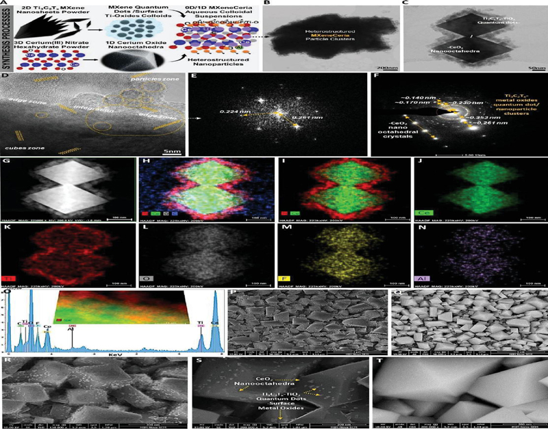
Figure 1 A, The synthesis procedures and B–T, physicochemical morphology and microstructural characterizations of MXeneCeria aqueous colloids.
3 Results and Discussions
Figure 1 depicts the schematic synthesis route as well as the morphology, microstructure, and elemental characterization of MXeneCeria. As shown in panel A, the TiCT MXene nanosheets were converted to quantum dots with surface titanium oxides through bath-sonication and homogenization for 60 and 30 min, respectively, followed by hydrothermal treatment of the suspensions at 180C for 14 hours. Further, these titanium–carbide/oxide quantum dots were integrated into the as-synthesized nano-octahedral particles of CeO (Supplementary Figure S1). The resultant 0D/1D heterostructure showed a unique morphology with high colloidal stability while retaining the main characteristics of parent materials (Supplementary Figures S2 and S3). Panels B and C display the transmission electron microscopy (TEM) of MXeneCeria clusters. As can be seen, these quantum dots are properly distributed into the ceria particles. Our high-resolution TEM image in panel D exhibits the integrated/lateral structure of the particles. The fast Fourier transform and selected-area-electron-diffraction images of MXeneCeria show the crystalline patterns and lattice d-spacing of its components (panels E, F).
Further, we performed a scanning-TEM (STEM) and energy-dispersive X-ray spectroscopy (EDX) area-scan and elemental-mapping analysis to confirm the purity of the material without significant detection of irrelevant chemical compositions (panels G–O). These data suggest a high level of structural integrity alongside typical elemental characteristics of these CeO and TiCT particles. In addition, panels P–T reveal different magnifications of scanning-EM (SEM) images of these aqueous colloids dried at room temperature. The SEM images captured from the surface of MXeneCeria at secondary-electrons (SE) mode and collective beam scanning (CBS) backscattered electrons illustrate its morphology/surface topography. These data suggest a uniform integration of metal-carbide/oxide particles of TiCT on the surface of lanthanide oxide ceria nano-octahedra.
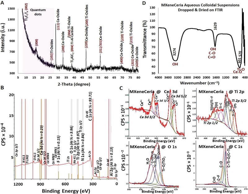
Figure 2 A: The XRD phase pattern. B,C: XPS wide and narrow scan fitting analysis. D: FTIR spectrum of MXeneCeria aqueous colloids.
We also performed X-ray diffraction and photoelectron spectroscopy (XRD/XPS) to characterize the phase pattern and surface chemistry of MXeneCeria. As shown in panel A of Figure 2, the XRD spectrum of this sample elaborates the detection of primary peaks associated with the chemical compositions of the synthesized composite heterostructure. Also, the XRD phase structure spectra of the raw and parent materials are represented in Supplementary Figure S4. Additionally, as shown in Figure 2B, C, the XPS survey spectra and narrow scan fitting analysis of the composing elements, including Ce 3d, O 1s, C 1s, and Ti 2p, suggest the successful synthesis and stable chemical bonding between different components of the heterostructured MXeneCeria. The detailed XPS components analysis as well as the corresponding narrow scan fittings of the F 1s, N 1s, and Al 2p are presented in Supplementary Figure S5 and Supplementary Tables S1 to S6. The Fourier transform infrared spectroscopy (FTIR) of this material suggest the presence of organic and/or inorganic components and bonds in its end-structure along with an enrichment of hydroxyl and fluorine-based surface functional groups (Figure 2D). These data align well with other properties of this new material and are in agreement with previous reports in the literature on the physicochemical characterizations of its parent TiCT MXene and CeO materials.
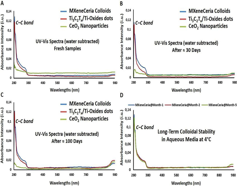
Figure 3 A–D: The UV–visible spectroscopic analysis of pure water, TiCT MXene nanosheets-derived quantum dots with surface metal oxides, CeO nanoparticles, and MXeneCeria at different wavelengths ranging from 280–980 nm. Data confirm the long-term durability and material stability of these aqueous colloidal suspensions at 4 C for up to around three months. The MXeneCeria showed no significant changes in the UV–Vis measurement from day 1 to day 100.
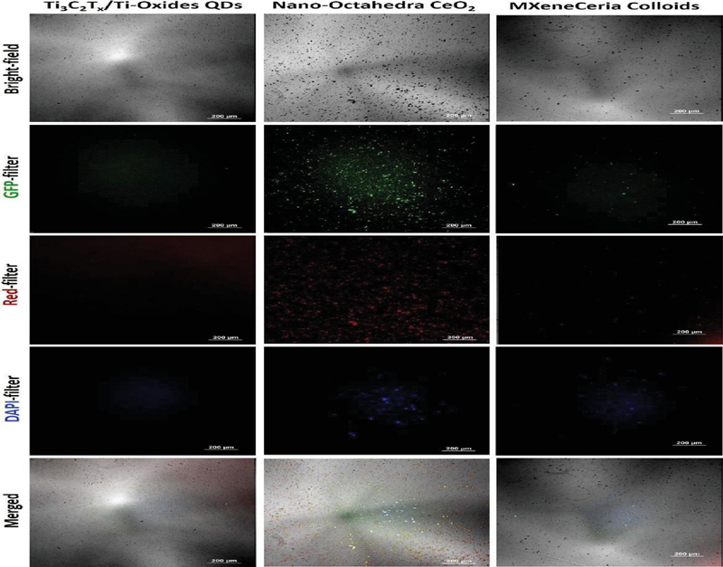
Figure 4 Detection of auto-fluorescence of TiCT MXene quantum dots with surface metal oxides, CeO nanoparticles, and MXeneCeria composite heterostructure at different wavelengths.
Furthermore, UV–visible spectroscopy (UV–Vis) was employed to measure the optical absorption and assess the enhanced colloidal dispersibility and stability of MXeneCeria in aqueous media. As demonstrated in Figure 3A–D, Our UV–Vis results suggest the optical absorption property and long-term stability of these collides at 4C for more than three months (around 100 days). The identification of C–C bonds in the UV–Vis spectra at a wavelength of 280 nm is a confirmation of the resistance of MXeneCeria against material decomposition in aqueous media.
Lastly, we have evaluated the auto-fluorescent properties of this colloidal material (Figure 4). Interestingly, MXeneCeria has shown fluorescence signals within different tested wavelengths, depicting its potential as a trackable nanoscale biomaterial for various diagnostics and nanomedicine approaches. As can be seen in Supplementary Figure S6, the aqueous colloidal dispersion of MXeneCeria showed relative auto-fluorescent properties, including excitation and emission across different regions and colors. While both of the parent materials used for developing MXeneCeria have been reported to possess high biocompatibility at controlled doses in their nature with intrinsic bioactivity properties for bio-related applications, future detailed studies should focus on investigating the biological/immunological activities and applications of MXeneCeria as well as the mechanisms behind it.
4 Conclusion
In this study, MXeneCeria is fabricated for the first time by integrating TiCT MXene nanosheet-derived quantum dots into the CeO nano-octahedral particles. This innovative 1D/0D assembly has been found to offer auto-fluorescent properties along with high dispersibility and stability for aqueous colloidal applications. Its microstructure may also benefit from high levels of biocompatibility and bioactivity properties of its parent materials, which is highly advantageous for bio-tracking applications. Another unique aspect of MXeneCeria composite heterostructure lies in the rational formulation of two immunomodulatory biomaterials in a single stable design. Together, these specifications pave the way for future studies.
Acknowledgments
The authors acknowledge research funding from the Natural Sciences and Engineering Research Council of Canada (RGPIN-2021–03951).
Competing Interests
The authors declare no competing interests.
CRediT Authorship Contribution Statement
Alireza Rafieerad: Conceptualization, Methodology, Investigation, Formal Analysis, Validation, Visualization, Writing – Original Draft, Writing – Review & Editing. Soofia Khanahmadi: Conceptualization, Formal Analysis, Writing – Original Draft, Writing – Review & Editing. Sanjiv Dhingra: Conceptualization, Methodology, Formal Analysis, Validation, Resources, Writing – Original Draft, Writing – Review & Editing.
Data Availability
The data that support the findings of this study are available from the corresponding author upon reasonable request.
References
[1] Dobrovolskaia, Marina A., and Scott E. McNeil. “Immunological Properties of Engineered Nanomaterials.” Nature Nanotechnology 2, no. 8 (2007): 469–78. https://doi.org/10.1038/nnano.2007.223.
[2] Zheng, Yuanyuan, Xiangqian Hong, Jiantao Wang, Longbao Feng, Taojian Fan, Rui Guo, and Han Zhang. “2D Nanomaterials for Tissue Engineering and Regenerative Nanomedicines: Recent Advances and Future Challenges.” Advanced Healthcare Materials 10, no. 7 (2021): 2001743. https://doi.org/10.1002/adhm.202001743.
[3] Chuang, Skylar T., Brandon Conklin, Joshua B. Stein, George Pan, and Ki-Bum Lee. “Nanotechnology-enabled immunoengineering approaches to advance therapeutic applications.” Nano Convergence 9, no. 1 (2022): 19. https://doi.org/10.1186/s40580-022-00310-0.
[4] Sang, Wei, Zhan Zhang, Yunlu Dai, and Xiaoyuan Chen. “Recent advances in nanomaterial-based synergistic combination cancer immunotherapy.” Chemical Society Reviews 48, no. 14 (2019): 3771–3810. https://doi.org/10.1039/C8CS00896E.
[5] Soleymaniha, Mohammadreza, Mohammad-Ali Shahbazi, Ali Reza Rafieerad, Aziz Maleki, and Ahmad Amiri. “Promoting role of MXene nanosheets in biomedical sciences: therapeutic and biosensing innovations.” Advanced healthcare materials 8, no. 1 (2019): 1801137. https://doi.org/10.1002/adhm.201801137.
[6] Rafieerad, Alireza, Weiang Yan, Ahmad Amiri, and Sanjiv Dhingra. “Bioactive and trackable MXene quantum dots for subcellular nanomedicine applications.” Materials & Design 196 (2020): 109091. https://doi.org/10.1016/j.matdes.2020.109091.
[7] Unal, Mehmet Altay, Fatma Bayrakdar, Laura Fusco, Omur Besbinar, Christopher E. Shuck, Süleyman Yalcin, Mine Turktas Erken et al. “2D MXenes with antiviral and immunomodulatory properties: A pilot study against SARS-CoV-2.” Nano Today 38 (2021): 101136. https://doi.org/10.1016/j.nantod.2021.101136.
[8] Rafieerad, Alireza, Weiang Yan, Keshav Narayan Alagarsamy, Abhay Srivastava, Niketa Sareen, Rakesh C. Arora, and Sanjiv Dhingra. “Fabrication of smart tantalum carbide MXene quantum dots with intrinsic immunomodulatory properties for treatment of allograft vasculopathy.” Advanced Functional Materials 31, no. 46 (2021): 2106786. https://doi.org/10.1002/adfm.202106786.
[9] Arora, Sumit, Jyutika M. Rajwade, and Kishore M. Paknikar. “Nanotoxicology and in vitro studies: the need of the hour.” Toxicology and applied pharmacology 258, no. 2 (2012): 151–165. https://doi.org/10.1016/j.taap.2011.11.010.
[10] Ernst, Lena M., and Victor Puntes. “How Does Immunomodulatory Nanoceria Work? ROS and Immunometabolism.” Frontiers in immunology 13 (2022): 750175. https://doi.org/10.3389/fimmu.2022.750175.
International Journal of Translational Science, Vol. 1, 351–362.
doi: 10.13052/ijts2246-8765.2024.044
© 2025 River Publishers