Age-Reversal: Advances in Regenerative Interventions
William Faloon
Co-Founder Life Extension® Group-Life Extension Foundation, 3600 West Commercial Boulevard, Fort Lauderdale, FL 33309, USA
E-mail: Bill@WilliamFaloon.com
Received 29 June 2026; Accepted 01 September 2026
Abstract
This perspective discusses emerging regenerative interventions that target biological aging. Over recent decades, scientific advances have expanded understanding of aging as a potentially modifiable biological process. Technologies under investigation include partial cellular reprogramming, senescent cell clearance, artificial-intelligence-driven drug discovery, organ regeneration, and modulation of epigenetic aging clocks.
Preclinical studies indicate that aged cells may regain more youthful functional characteristics under specific experimental conditions. For example, inhibition of interleukin-11 signaling initiated in late life extended median lifespan by approximately 20–25% in mice and partial reprogramming has improved tissue function and survival in aged mouse model. Developments in DNA-methylation-based biomarkers now permit estimation of biological age, allowing more efficient assessment of candidate interventions. All reprogramming data to date are preclinical, and human senolytic and IL-11 data are early-phase.
Although many findings remain experimental or early translational, they represent a convergence of approaches that may influence future biomedical research. This paper summarizes selected topics related to biological age modulation and highlights ongoing areas of investigation.
Keywords: Biological aging, cellular reprogramming, senolytics, epigenetic clock, regenerative medicine, longevity.
Abbreviations
| OSK: | Oct4, Sox2, Klf4 |
| OSKM: | Oct4, Sox2, Klf4, c-Myc (Yamanaka factors) |
| LNP: | lipid nanoparticles |
| PNP: | polymer nanoparticle |
| pDNA: | plasmid DNA |
| AI: | artificial intelligence |
| IL-11: | interleukin-11 |
Key Findings
Research into partial cellular reprogramming involves introducing genes associated with improved cellular function into aged cells, which may enable a shift toward a more youthful biological state in preclinical models.
Senolytic strategies aim to selectively eliminate senescent cells and have demonstrated potential to improve physical function and extend lifespan in experimental settings.
Aging involves multiple interconnected biological processes, suggesting that effective interventions may require combined approaches targeting gene regulation, inflammation, fibrosis, and cellular dysfunction.
Introduction
Advances in regenerative medicine and molecular biology have led to increasing interest in the possibility that biological aging may be modifiable. Earlier discussions of age reversal were often met with skepticism, particularly due to limited experimental evidence.
In recent decades, scientific progress has enabled further investigation into mechanisms that may influence biological aging. These include partial cellular reprogramming [1, 2], senescent cell clearance [3], artificial-intelligence-driven drug discovery [4, 5], organ regeneration [6], and interventions targeting epigenetic aging markers [7, 8].
While timelines for clinical application remain uncertain, current laboratory developments represent significant changes in the study of aging biology. This paper summarizes selected topics related to biological age modulation based on recent presentations and associated research.
Scope Statement. This is a narrative perspective. Topics were selected to reflect the author’s assessment of the most active areas of regenerative aging research; cited references were identified from PubMed and the author’s working knowledge of the field through 2026 and are illustrative rather than the product of a systematic search.
Cellular Reprogramming
Research into cellular reprogramming includes efforts to introduce genes associated with improved cellular function into aged cells and animal models. Findings suggest that aged cells may be driven toward a more youthful biological state [1] through the use of reprogramming factors identified by Yamanaka [9] (Figures 1 and 2).
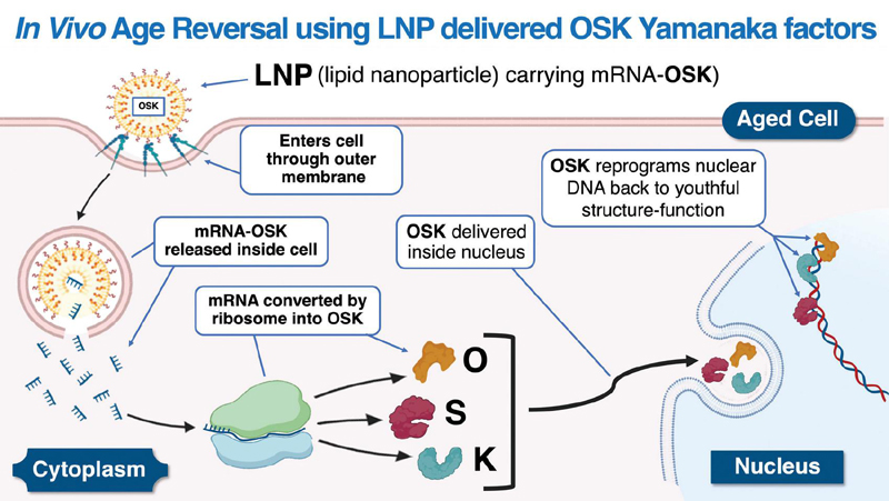
Figure 1 OSK RNA is encased in a lipid nanoparticle to enable it to cross the cell membrane. Once inside, the nanoparticle releases the OSK blueprint, which is translated into OSK proteins by ribosomes. These proteins pass through nuclear pores, bind nuclear DNA, and induce a reprogramming effect that improves markers of youthful structure and function in old cells [2].
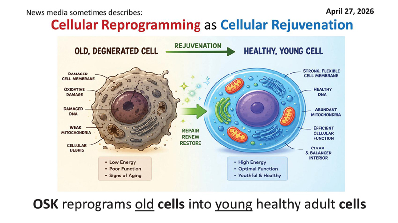
Figure 2 Reprogramming old cells into young cells.
These factors, commonly referred to as OSKM or OSK, demonstrate that differentiated cells can revert toward a more youthful condition under controlled experimental conditions [9]. Preclinical studies have reported improvements in tissue function, regenerative capacity, and survival in aged animal models following partial reprogramming [1, 2, 10]. A central goal of this field is the controlled rejuvenation of aged cells toward a more youthful, functional state without loss of cellular identity.
Tumorigenicity is the principal safety barrier to reprogramming-based rejuvenation, driven especially by c-Myc and by loss of cell-type identity during dedifferentiation. Cell-state-targeted strategies mitigate this: a Cdkn2a-promoter-driven OSK construct restricts reprogramming to aged and stressed cells and extended lifespan in naturally aged mice without altering tumor incidence [2].
Some research initiatives supported by longevity-focused organizations, including those affiliated with the Life Extension® group, are exploring delivery systems such as lipid and polymer nanoparticles to facilitate gene expression in vivo. These approaches remain at the preclinical or early translational stage (Figure 3).
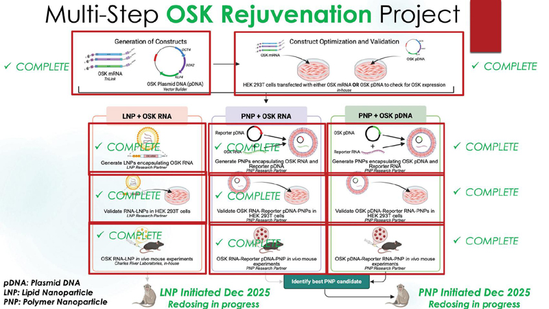
Figure 3 Flow chart describing the testing of LNP-OSK (lipid-nanoparticle-encased OSK), PNP-OSK (polymernanoparticle-encased OSK), and pDNA-OSK (plasmid-DNA-encased OSK) constructs first in vitro to validate efficacy, then in mice to validate systemic biodistribution, and with planned evaluation of the two most effective constructs (LNP-OSK and PNP-OSK) in aged nonhuman primates (see Table 1).
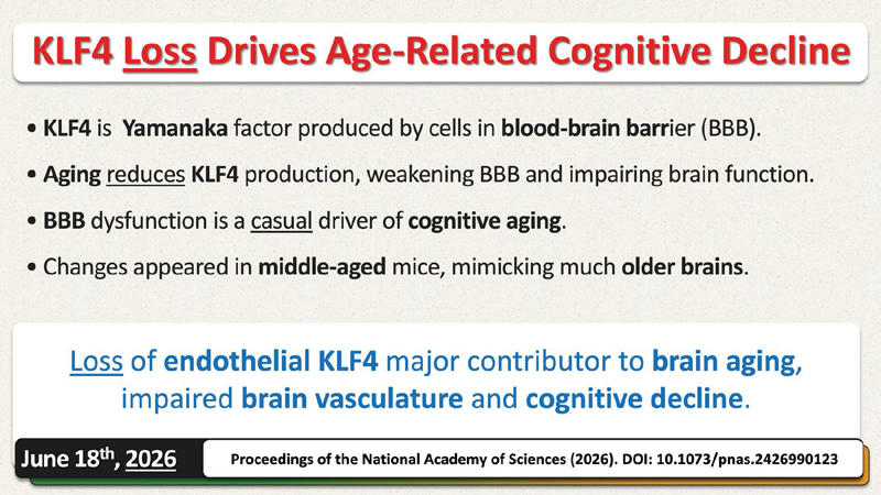
Figure 4 Endothelial KLF4 depletion and age-related neurovascular dysfunction, as reported in the Proceedings of the National Academy of Sciences [12].
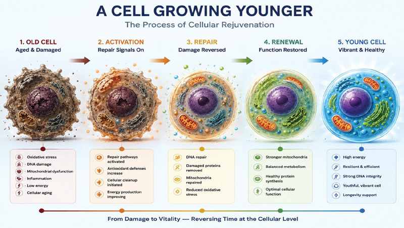
Figure 5 From damage to vitality – reversing time at the cellular level.
The epigenome – responsible for regulating gene expression – plays a central role in these processes [11]. Related work has also examined endothelial KLF4 depletion as a driver of age-related neurovascular dysfunction [12] (Figure 4).
Conceptually, these approaches aim to shift aged cells from a state of accumulated damage back toward youthful function (Figure 5).
Measurement of Biological Age
Efforts to quantify biological aging date back several decades. Early attempts were limited by available technology. However, advances since 2011 have enabled the development of DNA-methylation-based biomarkers capable of estimating biological age [13].
These epigenetic clocks allow researchers to evaluate changes in biological aging over shorter periods, rather than waiting for long-term lifespan outcomes. Such tools provide a practical method for assessing the impact of potential interventions.
Despite these advances, variability across individuals and uncertainty regarding clinical application remain important limitations.
Senescent Cells and Senolytics
Senescent cells are known to secrete inflammatory and tissue-altering factors that contribute to age-related dysfunction [14]. Their accumulation has been associated with conditions such as fibrosis, vascular disease, and frailty [15].
Senolytic strategies aim to selectively eliminate these cells. Compounds including fisetin, quercetin, and related agents have demonstrated beneficial effects in animal models, including improved physical function and lifespan extension [16, 17].
While early human investigations are ongoing, these interventions remain under evaluation for safety and effectiveness. These benefits are predominantly preclinical; human efficacy has not yet been established, and some human senolytic endpoints have been null.
Artificial Intelligence in Drug Discovery
Artificial intelligence is increasingly being applied to accelerate drug discovery processes [18]. Computational systems can identify potential therapeutic compounds and optimize molecular structures more efficiently than traditional methods [4, 5].
Organizations supported in part by early-stage longevity-focused funding have contributed to this area, with AI-driven drug development collaborations advancing candidate therapies toward the clinic [4, 5]. These approaches may be particularly relevant to aging research given the complexity of interacting biological pathways.
Table 1 Summary of aging interventions and representative references
| Approach | Mechanism | Evidence | Stage | References |
| OSK reprogramming | Epigenetic reset | Animal studies | Preclinical | [1, 2, 10] |
| Senolytics | Cell clearance | Animal + early human | Early clinical | [16, 17] |
| AI drug discovery | Target identification | Ongoing | Translational | [4, 5] |
| IL-11 inhibition | Anti-fibrotic | Animal | Early clinical | [24] |
Table 2 Interleukin-11 (IL-11) as a longevity and anti-fibrotic target
| Aspect | Finding | Reference |
| Biological role | IL-11 is a mediator of fibrotic signaling and contributes to age-related tissue dysfunction | [22, 23] |
| Preclinical effect | Inhibition of IL-11 signaling extends mammalian healthspan and lifespan, including when initiated in later life | [24] |
| Translational status | IL-11 inhibition is under early clinical evaluation; human efficacy remains to be established | [24–26] |
Anti-Fibrotic Strategies and IL-11
Fibrosis contributes to organ dysfunction across multiple age-related conditions [19–21]. Interleukin-11 has been identified as a mediator in fibrotic signaling pathways [22, 23].
Animal studies have reported that inhibition of IL-11 can improve healthspan and survival when administered in later life stages [24]. These findings have attracted research and development interest, including investment and early clinical evaluation [25, 26] (Table 2).
While these results are promising, IL-11 inhibition remains in early clinical evaluation, and efficacy in humans has not yet been established (Table 2).
Research Funding and Scientific Expansion
In recent years, there has been increased investment in longevity and regenerative medicine research. This includes funding from private organizations, biotechnology firms, and institutional research groups. Substantial capital from the technology and venture-investment sectors has been directed toward longevity research, and several longevity-focused startups have experienced rapid increases in valuation, underscoring the intensifying commercial interest in this field.
Many of these efforts distinguish between extending lifespan and improving healthspan, emphasizing preservation of function and independence. Rather than treating cancer, dementia, heart disease, and frailty as separate phenomena, an increasing number of researchers and institutions recognize that most age-related chronic diseases are downstream consequences of shared aging biology [27]. This shift reflects a broader focus on addressing underlying aging mechanisms rather than isolated diseases.
Research initiatives affiliated with organizations such as Life Extension® have contributed to this evolving landscape alongside academic and commercial efforts.
Systemic Interventions
Aging involves multiple interconnected biological processes, suggesting that effective interventions may require combined or systemic approaches. Current research is exploring strategies that simultaneously address gene regulation, inflammation, fibrosis, and cellular dysfunction.
Advances in delivery technologies and biological understanding are expanding the range of potential interventions. However, challenges related to safety, scalability, and long-term outcomes remain.
Limitations
These findings carry important limitations. Partial reprogramming risks tumorigenesis and loss of cell identity; human senolytic and IL-11 trials remain early-phase with efficacy unestablished, and some human senolytic endpoints have been null; and epigenetic aging clocks show inter-individual variability and unresolved questions of causal validity. Much of the cited evidence derives from animal models, and translation to human healthspan is not assured. Life Extension® will be funding murine xenograft studies of four common human cancer cell lines to assess if robust in vivo enhancement of OSK transfection to nuclear DNA results in tumor expansion, tumor shrinkage, or no change compared to control animals. If there are no changes in tumor mass in this study, it will corroborate two previous published studies showing no increased cancer risk in OSKM and OSK treated mice. Both of these previous studies demonstrated age reversal effects.
Conclusion
Advances in regenerative biology, molecular science, and computational technologies are contributing to a growing body of research focused on biological aging.
While preclinical findings suggest that aspects of aging may be modifiable, substantial uncertainties remain regarding clinical translation. Continued investigation will determine whether these approaches can be safely and effectively applied to improve human healthspan.
Ongoing Unpublished Work (Context Only)
An internal program is testing LNP-OSK (lipid-nanoparticle-encased OSK), PNP-OSK (polymer-nanoparticle-encased OSK), and pDNA-OSK (plasmid-DNA-encased OSK) constructs in vitro to validate efficacy, then in mice to validate systemic biodistribution, with the two most effective constructs planned for infusion into aged nonhuman primates. No results are yet available; this design is presented for context only and does not constitute evidence.
Conflict of Interest
The author, William Faloon, is Co-Founder of the Life Extension Group, a nonprofit organization that funds and conducts research in longevity and regenerative medicine. Several interventions discussed in this article, including lipid-nanoparticle, polymer-nanoparticle-, and plasmid-DNA-delivered OSK reprogramming constructs and the associated primate study, are part of research programs supported by the Life Extension Foundation and affiliated entities. The author has a financial and organizational affiliation with programs investigating OSK-based interventions; readers should interpret discussion of these approaches in that context.
Funding
This work reflects perspectives informed by research initiatives supported in part by longevity-focused organizations. No specific grant was designated for this manuscript.
Acknowledgement
The author gratefully acknowledges Mahwish Aurangzeb, M.D., MPH, Medical Editor at Life Extension Magazine, for editorial development, manuscript preparation, and medical review of this work.
References
[1] A. Ocampo et al., “In vivo amelioration of age-associated hallmarks by partial reprogramming,” Cell, vol. 167, no. 7, pp. 1719–1733, Dec. 15, 2016.
[2] S. K. Sahu et al., “Targeted partial reprogramming of age-associated cell states improves markers of health in mouse models of aging,” Sci. Transl. Med., vol. 16, no. 764, p. eadg1777, Sep. 11, 2024.
[3] S. Chaib, T. Tchkonia, J. L. Kirkland, “Cellular senescence and senolytics: The path to the clinic,” Nat. Med., vol. 28, no. 8, pp. 1556–1568, Aug. 2022.
[4] S. Arora et al., “Discovering geroprotectors through the explainable artificial intelligence-based platform AgeXtend,” Nat. Aging, vol. 5, no. 1, pp. 144–161, Jan. 2025.
[5] H. Li et al., “Artificial intelligence across the aging continuum: Mechanistic geroscience, therapeutic innovation, and clinical impact,” Ageing Res. Rev., vol. 120, p. 103205, Jun. 10, 2026.
[6] J. H. Elisseeff, S. F. Badylak, J. D. Boeke, “Immune and genome engineering as the future of transplantable tissue,” N. Engl. J. Med., vol. 385, no. 26, pp. 2451–2462, Dec. 23, 2021.
[7] A. A. Johnson, D. A. Sinclair, “Turning back time: A comprehensive list of interventions that decrease nextgeneration epigenetic aging clocks in humans,” Front. Genet., vol. 17, p. 1836446, 2026.
[8] K. N. Fitzgerald et al., “Potential reversal of epigenetic age using a diet and lifestyle intervention: A pilot randomized clinical trial,” Aging (Albany NY), vol. 13, no. 7, pp. 9419–9432, Apr. 12, 2021.
[9] K. Takahashi, S. Yamanaka, “Induction of pluripotent stem cells from mouse embryonic and adult fibroblast cultures by defined factors,” Cell, vol. 126, no. 4, pp. 663–676, Aug. 25, 2006.
[10] C. C. Macip et al., “Gene therapy-mediated partial reprogramming extends lifespan and reverses age-related changes in aged mice,” Cell Reprogram., vol. 26, no. 1, pp. 24–32, Feb. 2024.
[11] W. Zhang et al., “The ageing epigenome and its rejuvenation,” Nat. Rev. Mol. Cell Biol., vol. 21, no. 3, pp. 137–150, Mar. 2020.
[12] M. Dhar et al., “Endothelial KLF4 depletion drives age-related neurovascular dysfunction,” Proc. Natl. Acad. Sci. U.S.A., vol. 123, no. 25, p. e2426990123, Jun. 23, 2026.
[13] M. E. Levine et al., “An epigenetic biomarker of aging for lifespan and healthspan,” Aging (Albany NY), vol. 10, no. 4, pp. 573–591, Apr. 18, 2018.
[14] J. L. Kirkland, T. Tchkonia, “Cellular senescence: A translational perspective,” EBioMedicine, vol. 21, pp. 21–28, Jul. 2017.
[15] J. Kaur, J. N. Farr, “Cellular senescence in age-related disorders,” Transl. Res., vol. 226, pp. 96–104, Dec. 2020.
[16] D. E. Forman et al., “Impact of geroscience on therapeutic strategies for older adults with cardiovascular disease,” J. Am. Coll. Cardiol., vol. 82, no. 7, pp. 631–647, Aug. 15, 2023.
[17] P. D. Robbins et al., “Senolytic drugs: Reducing senescent cell viability to extend health span,” Annu. Rev. Pharmacol. Toxicol., vol. 61, pp. 779–803, Jan. 6, 2021.
[18] K. Huanbutta et al., “Artificial intelligence-driven pharmaceutical industry: A paradigm shift,” Eur. J. Pharm. Sci., vol. 203, p. 106938, Dec. 1, 2024.
[19] M. Selman, A. Pardo, “Fibroageing: An ageing pathological feature,” Ageing Res. Rev., vol. 70, p. 101393, Sep. 2021.
[20] L. L. Ren et al., “TGF-beta as a master regulator of aging-associated tissue fibrosis,” Aging Dis., vol. 14, no. 5, pp. 1633–1650, Oct. 1, 2023.
[21] D. C. Rockey, P. D. Bell, J. A. Hill, “Fibrosis – a common pathway to organ injury and failure,” N. Engl. J. Med., vol. 372, no. 12, pp. 1138–1149, Mar. 19, 2015.
[22] S. Schafer et al., “IL-11 is a crucial determinant of cardiovascular fibrosis,” Nature, vol. 552, no. 7683, pp. 110–115, Dec. 7, 2017.
[23] S. A. Cook, S. Schafer, “Hiding in plain sight: Interleukin-11 as a master regulator of fibrosis,” Annu. Rev. Med., vol. 71, pp. 263–276, Jan. 27, 2020.
[24] A. A. Widjaja et al., “Inhibition of IL-11 signalling extends mammalian healthspan and lifespan,” Nature, vol. 632, no. 8023, pp. 157–165, Aug. 2024.
[25] S. A. Cook, “Understanding interleukin-11 as a disease gene and therapeutic target,” Biochem. J., vol. 480, no. 23, pp. 1987–2008, Dec. 13, 2023.
[26] B. Ng et al., “Interleukin-11 is a therapeutic target in idiopathic pulmonary fibrosis,” Sci. Transl. Med., vol. 11, no. 511, Sep. 25, 2019.
[27] S. B. Kritchevsky, S. R. Cummings, “Geroscience: A translational review,” JAMA, vol. 334, no. 12, pp. 1094–1102, Sep. 23, 2025.
International Journal of Translational Science, 2026, 159–170
doi: 10.13052/ijts2246-8765.2026.007
© 2026 River Publishers