Combining Senolytic, Stem Cell, and NAD+ Strategies as an Integrated Protocol
Based on a lecture delivered at the HEALinc Future Health Innovation Summit, May 2026.
Gregory Kelly
Qualia Life Sciences, 2011 Palomar Airport Road, Suite 101 – PMB 160, Carlsbad, CA 92011, USA
E-mail: greg@qualialife.com
Received 30 June 2026; Accepted 06 August 2026
Abstract
Human life expectancy has risen faster than the span of life spent in good health, producing a widening healthspan–lifespan gap that now approaches a decade globally and exceeds 12 years in the United States. Conceptual approaches for aging intervention range from largely mechanistic-based frameworks, such as the hallmarks of aging, to more function-first approaches like the World Health Organization’s intrinsic capacity model. Because biological aging is driven not by a single mechanism, but by a network of interacting hallmarks, and because many aspects of physical and cognitive function can be declining together leading up to the time period spent in the healthspan–lifespan gap, interventions that target only one process or area of function are unlikely to compress this gap substantially. This perspective makes the translational case for combining – and deliberately sequencing – potentially complementary categories of intervention: senolytics that clear senescent cells, regenerative support that supports stem cell function, joint support to enhance mobility, and nicotinamide adenine dinucleotide (NAD+) repletion that supports cellular energetics. The rationale rests on documented cross-talk among these mechanisms: as an example, senescent cells consume NAD+ and impair the niches in which stem cells operate, so clearing senescent burden plausibly creates a more permissive environment for both NAD+ restoration and regenerative signaling. A recently published preclinical study demonstrated synergistic healthspan and lifespan extension when a senolytic immunotherapy was combined with personalized mesenchymal stem cells in accelerated-aging models, providing proof of concept for the combination principle. A 12-week pilot trial using newer generation methylation clocks (e.g., DunedinPACE, OMICmAge) is planned to test an integrated nutraceutical and botanical protocol directly.
Keywords: Healthspan, NAD+, senolytic, stem cells.
Notations and Abbreviations
| CXCL9: | C-X-C motif chemokine ligand 9 |
| CXCL10: | C-X-C motif chemokine ligand 10 |
| CXCL11: | C-X-C motif chemokine ligand 11 |
| CCL8: | C-C motif chemokine ligand 8 |
| CXCL8: | C-X-C motif chemokine ligand 8; also known as IL-8 (interleukin 8) |
| DNA: | deoxyribonucleic acid |
| GDF-11: | growth differentiation factor 11 IL-1 (interleukin-1 beta) |
| IL-6: | interleukin-6 IL-11 (interleukin-11) |
| NAD+: | nicotinamide adenine dinucleotide |
| SASP: | senescence-associated secretory phenotype |
| TNF: | tumor necrosis factor |
| VEGF: | vascular endothelial growth factor |
| VEGFA: | vascular endothelial growth factor A) |
| WHO: | World Health Organization |
| YKL-40: | also called Chitinase-3-like protein 1 (CHI3L1) |
1 The Healthspan–Lifespan Gap as the Central Problem
For most of the modern era, the central achievement of medicine and public health has been the extension of life. That achievement is real, but it has outpaced a second, arguably more important goal: extending the years lived in good health. The difference between how long people live and how long they live well is the healthspan–lifespan gap, and it has been widening.
The most comprehensive recent quantification comes from an analysis of World Health Organization (WHO) data covering 183 member states over two decades. Globally, the gap between life expectancy and health-adjusted life expectancy widened to 9.6 years. The United States fares worse than the global average on every measure. Over the past two decades the mean US healthspan–lifespan gap grew from 10.9 to 12.4 years – 29% wider than the global mean – and the gap among American women widened from 12.2 to 13.7 years, 2.6 years beyond that of men and 32% above the global mean for women (Figure 1A) [1]. US life expectancy rose over the period (from 79.2 to 80.7 years in women and from 74.1 to 76.3 years in men), while health-adjusted life expectancy barely moved – unchanged in women and up just 0.6 years in men (Figure 1B) [1]. Yes, life expectancy has increased, but the added years of life gained in the US over the past two decades have been accruing almost entirely as years of compromised health. These are not abstract figures. They describe well over a decade, near the end of most lives, lived with meaningful limitations.
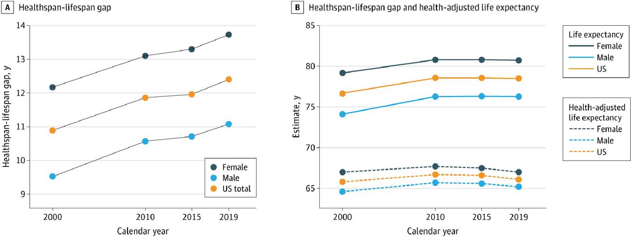
Figure 1 (A) The US healthspan–lifespan gap with stratification by sex. (B) US life expectancy and health-adjusted life expectancy with stratification by sex. Reproduced from A. Garmany and A. Terzic, “Global healthspan-lifespan gaps among 183 World Health Organization Member States,” JAMA Netw Open, vol. 7, no. 12, p. e2450241, 2024 Dec 2, doi:10.1001/jamanetworkopen.2024.50241. License: CC BY 4.0 (https://creativecommons.org/licenses/by/4.0/).
This reframes the objective of longevity medicine. The goal is not simply to add years to life but to add life to years – to compress the period of morbidity so that the function is preserved closer to the full span of a person’s lifespan. Achieving that requires understanding why function declines in the first place, and that understanding has matured considerably over the past 15 years.
2 From Hallmarks to Physical and Cognitive Function
The dominant conceptual framework for the biology of aging is the hallmarks of aging: a set of interrelated cellular and molecular processes whose progressive dysregulation drives age-related decline. Originally enumerated as nine [2], the framework has since expanded to 12 hallmarks with the addition of disabled macroautophagy, chronic inflammation, and dysbiosis [3]. These hallmarks are grouped as primary drivers of damage, antagonistic responses that become harmful with age, and integrative consequences that manifest at the level of the whole organism [3, 4]. More recently, two additional hallmarks have been proposed: (1) extracellular matrix changes, and (2) psychosocial isolation [5]. The addition of the latter acknowledged that social and behavioral factors are an essential determinant of aging with a wide-ranging impact on physical and cognitive function.
Telomere attrition, genomic instability, epigenetic alteration, loss of proteostasis, mitochondrial dysfunction, cellular senescence, stem cell exhaustion, altered intercellular communication, and chronic inflammation are among the processes now understood to act not in isolation but as a densely interconnected web [3]. Because the hallmarks are an interconnected network, intervening in one hallmark may impact others to a degree, and some interventions – calorie restriction as an example [6] – can modestly impact multiple hallmarks.
Another practical implication of this interconnection implies that an intervention aimed at a single hallmark may address only one node in a network whose other nodes continue to drive decline. If senescent cells accumulate while stem cell pools are depleted and cellular energy metabolism falters, correcting any one of these alone leaves the others unmanaged and able to perpetuate dysfunction. This is the foundational argument for combination approaches, and the remainder of this paper develops it.
Yet hallmarks alone do not tell us what to measure or what patients actually experience leading up to, and throughout the growing healthspan–lifespan gap. A complementary framework, advanced by the WHO and increasingly linked to geroscience, is intrinsic capacity: the composite of an individual’s physical and mental capacities, spanning locomotion, cognition, psychological well-being, vitality, and sensory function. A recent narrative review proposed that intrinsic capacity can serve both as a measurable indicator of global function and as a target for geroscience interventions, mapping specific hallmarks of aging onto each functional domain (Figure 2) [7]. This shift – from disease and from molecular abstraction toward measurable function – matters because function is what people value and what ultimately defines healthspan.
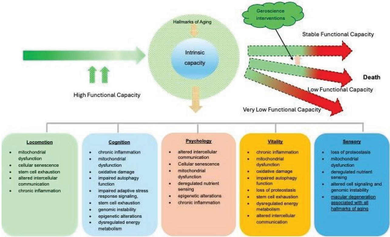
Figure 2 Mechanism linking the hallmarks of aging with decline in intrinsic capacity. Reproduced from S. Zhu, L. Tao, C. Fan, Y. Wei, J.K. Chhetri, and P. Chan P, “Aligning intrinsic capacity and geroscience: linking function with biology,” Aging Dis., January 13, 2026. doi:10.14336/AD.2025.1419. License: CC BY 4.0 (https://creativecommons.org/licenses/by/4.0/).
A provocative extension of the function-first view places the brain at the center. In an editorial advancing a “brain-first” systems framework, Lakhan argued [8] that progressive disruption of neural networks governs functional decline across multiple physiological systems regardless of peripheral biological age, and that when brain function deteriorates, lifespan may persist while meaningful healthspan collapses. As an editorial, this is a proposed conceptual framework rather than an empirical finding, but it captures an intuition consistent with the intrinsic-capacity view: the value of additional years depends on the preservation of the capacities – cognitive, autonomic, affective – that allow those years to be experienced fully. This “brain-first” framework can be tied back to the hallmarks of aging, since, for example, neuroinflammation and brain energetics link the brain-first and hallmarks of aging perspectives.
Taken together, these frameworks point in one direction: aging is a multi-mechanism, function is the endpoint that matters, and preserving function may require intervening across multiple mechanisms or systems at once to globally impact many areas of physical and cognitive performance.
3 Two Different Objectives: Slowing Aging and Repairing Damage
If combination intervention is to be implemented it is worth distinguishing the kinds of interventions that might be combined. Pfleger proposed [9] a useful framing that divides the longevity field into two broad strategies. The first is slowing aging: continuous or frequent interventions – caloric restriction, exercise, and related approaches – that reduce the rate at which damage accumulates. The second is damage repair or rejuvenation: intermittent interventions – such as senolytics and stem cell therapies – that are aimed at rejuvenation or reversing damage already present [9].
The distinction carries a practical consequence. Slow-aging therapies generally have their largest effects when started early, because they act on the rate of future damage; they do comparatively little for those already near the end of their lifespans. Rejuvenation interventions, by contrast, can in principle benefit those who are already old, because they address damage that has already accumulated [9].
To implement an approach based on this framing, it is important to determine what therapies fall into which strategy. Slow-aging strategies started too late in a person’s life may be relatively ineffective in narrowing the healthspan–lifespan gap. Rejuvenation therapies started far too early may be ineffective, or of lesser impact because not much damage has accumulated. In most lives there is likely to be a prolonged period of time where focusing on both strategies simultaneously will have an impact.
This is the conceptual hinge of the present argument. Meaningfully impacting the healthspan–lifespan gap may require addressing multiple forms of accumulated damage simultaneously, in combination with other interventions focused on slowing aging in a population within an age or functional health range that is still likely to benefit from both.
An example approach would be a protocol that offers support for senescent cell clearance and stem cell activity – rejuvenation strategies – while simultaneously helping to manage inflammatory, structural and mitochondrial functions – slow-aging strategies. What remains is to determine whether strategies based upon these different mechanisms genuinely interact – that combining them is additive – and whether the order in which they are deployed matters.
4 The Mechanistic Case for Combining – and Sequencing
The strongest justification for a combined protocol is that its components are not independent. Senescent cells (and their senescence-associated secretory phenotype [SASP] characterized by cytokine signaling molecules), NAD+ metabolism, and stem cell function are bound together by documented cross-talk, and that cross-talk implies a logic for sequencing as well as for combination.
Senescent cells and NAD+ are metabolically entangled, with cellular senescence and NAD+ metabolism interacting bidirectionally. Cellular senescence is triggered by three broad categories of issues: (1) replicative senescence resulting from telomere attrition, (2) premature stress-induced senescence caused by factors such as mitochondrial dysfunction, DNA damage, nutrient inadequacy, oxidative stress, proteolytic stress, and specific therapies that are intended to cause cellular stress (so-called therapy-induced senescence), and (3) secondary or paracrine senescence – the process by which a primary senescent cell spreads its state of senescence to healthy, neighboring cells via SASP [10]. Low intracellular NAD+ can make a cell less resistant to many of these insults. At the same time, senescent cells and their SASP activate NAD+-consuming enzymes, depleting tissue NAD+. This combination – low NAD+ increasing susceptibility to cellular senescence and senescent cells contributing to NAD+ depletion – may be a physiological viscous cycle contributing to age-related functional decline [11].
The interaction between cellular senescence and NAD+ is further complicated, because NAD+ appears to be necessary for senescent-cell function and SASP production. This could mean that replenishing NAD+ in the presence of a high senescent-cell burden could in principle feed some of the processes contributing to inflammatory signaling and secondary senescence [11]. This relationship is why sequence may matter. Reducing senescent burden first, then focusing on NAD+ repletion, is mechanistically more defensible than interventions aimed at increasing NAD+ into an unmanaged senescent landscape. Depletion of senescent cells has been shown to partially recover NAD+ levels in aging tissues, suggesting that the two interventions, properly ordered, may reinforce one another. Because of the interactions between cellular senescence and NAD+, Chini et al. have proposed [11] combining senolytics with NAD+ replenishment as a way to promote healthier aging (Figure 3).
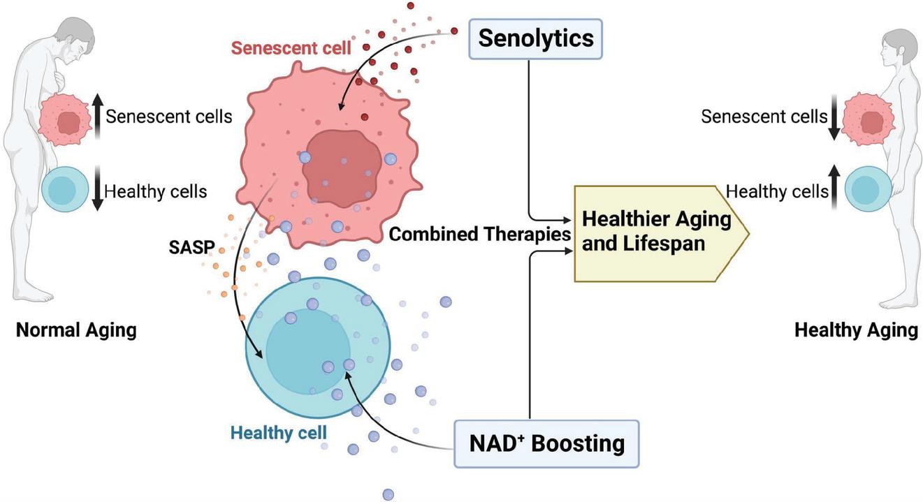
Figure 3 Theoretical framework for combined administration of senolytics and NAD+ boosting (replacement) therapies to promote healthier aging and lifespan. Reproduced from C.C.S. Chini, H.S. Cordeiro, N.L.K. Tran, and E.N. Chini, “NAD metabolism: role in senescence regulation and aging,” Aging Cell, vol. 23, no. 1, p. e13920, 2024, doi:10.1111/acel.13920. License: CC BY 4.0 (https://creativecommons.org/licenses/by/4.0/)
The senescent environment impairs stem cell function. Stem cell exhaustion is itself a hallmark of aging, but a comprehensive review of stem cell aging emphasizes that aged stem cells are shaped not only by intrinsic damage but by an aged, inflamed environment and stem cell niche. The accumulation of senescent cells in the stem cell niche can negatively affect stem cell behavior through paracrine factors, and – importantly – these effects could be substantially reversible if the environment were improved, for example, by reducing inflammation or eliminating senescent cells [12]. The niche, in other words, can hold otherwise-functional stem cells in a dysfunctional state.
This has immediate implications for regenerative strategy. An in vitro study modeling young versus aged hematopoietic stem cell niches found that the aged niche environment altered the development and inflammatory profile of bone-marrow-derived dendritic cells, and the authors advanced this as a likely explanation for why heterochronic stem cell transplantation has often underperformed expectations: the problem may lie in the aged niche rather than in the transplanted cells themselves [13]. Consistent with this, transplanting adipose-derived mesenchymal stem cells from old donors – but not from young donors – was shown to induce physical dysfunction in older recipient mice, with single-cell analysis revealing a naturally occurring senescent-like population within the old-donor cells [14]. Both findings point to the same lesson: introducing regenerative cells into, or deriving them from, a senescent context can blunt or even reverse the intended benefit.
Clearing senescence makes regeneration feasible – senescent-cell clearance and regenerative support are natural partners. In the context of osteoarthritis, a field where both senescence and impaired repair are central, it has been proposed that senolytics and mesenchymal stem cells could be used together to synergistically amplify their anti-inflammatory and regenerative effects – senolytics clearing senescent cells from the host (and removing any harmful senescent cells among the therapeutic population), while stem cells repair damaged tissue [15]. In the hematopoietic compartment, aging has been attributed substantially to the clonal accumulation of a senescent cell cluster, and clearance of that population with senotherapy has been described as feasible and potentially beneficial in humans [16].
The mechanistic picture that emerges is as follows: senescent cells consume NAD+ and degrade the niches stem cells depend on; clearing them plausibly relieves both burdens at once, creating conditions under which NAD+ repletion and regenerative support can act more effectively. This is the biological argument for a sequenced protocol: clear first, then rebuild and re-energize.
5 Assembling the Pieces
A mechanistic rationale, however coherent, is not evidence that combination works. That evidence has begun to arrive. In June 2026, a study published in the Journal of Translational Medicine tested the combination principle directly in animal models of accelerated aging [17].
The investigators combined a senolytic immunotherapy – SenoVax, a dendritic-cell-based approach targeting senescent-cell antigens – with personalized, pluripotent-stem-cell-derived mesenchymal stem cells, using two established models: carbon-tetrachloride-induced liver injury and doxorubicin-induced systemic senescence. Across both models, the combination outperformed either monotherapy, producing synergistic improvements in biochemical markers of liver function, reductions in senescence-associated inflammatory mediators (including IL-11, YKL-40 [CHI3L1], and IL-6), restoration of regenerative signaling factors (Klotho, VEGF, GDF-11), improved physical performance, and – most strikingly – extended survival. Senolytic monotherapy yielded partial improvement, while stem cells alone showed limited activity in the presence of a high senescent-cell burden [17].
That last detail is the empirical echo of the mechanistic argument in Section 4: stem cells underperform when the senescent environment is left intact, and pre-clearing senescent cells could unlock their regenerative potential. The authors interpreted their results as support for a model in which senescent cells and their SASP directly suppress stem-cell-mediated repair, and in which senolytic therapy acts as an adjuvant that makes regenerative therapy work.
Two caveats are essential for honest translation. First, these were models of accelerated aging induced by toxic and chemotherapeutic insults, not natural aging; the authors explicitly acknowledge this limitation and note that studies in natural aging are underway. Second, the specific agents tested – a dendritic-cell immunotherapy and engineered stem cells – are not the botanical formulations discussed below; what transfers is the principle of senolytic-plus-regenerative synergy, not the particular interventions. With those caveats, the study is a meaningful proof of concept: in living animals, combining senescent-cell clearance with regenerative support produced benefits that neither approach achieved alone.
6 Preliminary Studies on Botanical and Nutraceutical Formulas
Translating the combination principle into accessible human interventions is the work my colleagues and I are pursuing through a series of botanical and nutraceutical formulations, each targeting one node of the network, designed to be used in sequence. The clinical evidence is preliminary – a peer-reviewed proteomics pilot, two placebo-controlled NAD+ studies, and small open-label pilot studies – and is presented here as a directional signal rather than confirmation.
NAD+ declines with age across tissues, though it is consistently higher in lifelong athletes than in untrained individuals – evidence that the decline is to some extent modifiable though not completely preventable [18]. Two double-blind, placebo-controlled studies of a multi-ingredient NAD+ formulation, in which participants were assigned to groups by a systematic (non-random) stratified allocation, tested whether this decline can be reversed. In the first (, aged 40–65), active treatment raised whole-blood NAD+ by an average of 74% versus 4% with placebo (), with improvements in aging-symptom scores limited to males [19]. In the second and larger study (, aged 35–76), NAD+ rose 67% in the active intervention group versus 4% with placebo (), and quality-of-life gains were broader: emotional well-being and vitality improved across participants, with aging-symptom improvements in females [20]. Because allocation was not randomized, these results are more susceptible to baseline imbalance and should be interpreted accordingly.
Joint function is an instructive target for intervention because it is one of the leading contributing factors to the healthspan–lifespan gap. It is central to locomotion, one of the five domains of intrinsic capacity, WHO’s function-centered framework. Mobility restriction is also one of the core drivers of psychosocial isolation, a newly added hallmark of aging. Lastly, structural and functional changes with joints begin sooner than most people expect.
Mitochondrial performance throughout the body, including in tendons, ligaments, cartilage, and synovial membranes, begins to decline as early as the fourth decade of life, reducing the energy available for producing the structural (e.g., collagen, proteoglycans) and lubricating (e.g., hyaluronic acid, lubricin) molecules that maintain joint tissues. The consequences are detectable remarkably early: in a population-based birth cohort, structural knee abnormalities on MRI were already frequent at age 33, with small articular cartilage lesions identified in 56.2% of patellofemoral and 25.3% of tibiofemoral joints among a largely asymptomatic population [21]. Structural and functional decline in joints, in other words, is an early and near-universal manifestation of the same mitochondrial and senescent processes that drive systemic aging – making it both a sensible early target and a useful readout.
With this in mind, we have conducted two studies focused on joint function. The first was an open-label study of a joint-health formulation – active ingredients included an eggshell membrane extract to supply collagen, elastin, and hyaluronic acid, L-carnitine for mitochondrial support, and a blend of botanical ingredients including extracts of boswellia, cucumber, ginger, tamarind, and turmeric. Thirteen healthy adults aged 36 to 62 took the formula for 28 days. There were substantial improvements across joint function measures over 28 days, including a 58% improvement in overall score and a 66% reduction in scores related to how occasional stiffness or discomfort in knees or low back impacted the ability to be comfortable throughout the day in a variety of activities, staying active and sleeping well [22].
A second study (, aged 35–79) used a senolytic formula containing a blend of plant extracts standardized for polyphenols (e.g., fisetin, quercetin, luteolin, oleuropein, isoflavones, curcumin, silymarin) and piperlongumine, as well as a proprietary extract of Panax notoginseng and Rosa roxburghii. The study was placebo-controlled and used an intermittent dosing approach – 2 days of the senolytic formula, followed by 12 days with no intervention. The 2-day dosing cycle was completed three times over about a 5-week time span. Joint health and performance scores had a significantly greater change from baseline in the senolytic group compared to placebo (66% versus 39%, ) [23].
The same senolytic formula was also used in a proteomics pilot study using Olink’s 45-cytokine panel. In this peer-reviewed, decentralized single-arm pilot ( analyzed, aged 45–79), two days of the senolytic blend did not significantly change the four pre-specified primary SASP biomarkers (TNF, IL-1, CXCL8, VEGFA). Exploratory, unadjusted analyses did, however, reveal reductions in the senescence-associated chemokines CXCL9, CXCL10, CXCL11, and CCL8 – signals consistent with reduced SASP-associated inflammation, but which did not survive correction for multiple comparisons and therefore remain hypothesis-generating. Quality-of-life measures improved, though the single-arm design makes these expectancy-sensitive [24].
7 An Integrated Protocol and the Path Forward
The mechanistic rationale and the preliminary signals have converged on a specific, testable protocol that respects the sequencing logic developed above. Rather than administering all components simultaneously, the protocol front-loads a senescent cell intervention, then introduces stem cell (regenerative) support, both of which follow an intermittent administration approach. NAD+ repletion and connective-tissue/inflammatory signaling support are provided on a more continuous basis. The proposed protocol is as follows:
• Days 1–2: senolytic compounds, to manage senescent-cell burden first;
• Days 4–7: stem-cell-activating compounds, introduced into the now more permissive environment;
• Days 3–30: an NAD+-replenishing formulation supplying NAD+ precursors and cofactors once senescent demand has been reduced;
• Days 3–30: botanical extracts and extracellular-matrix biomolecules supporting joint and connective tissue.
This sequence is the practical expression of biology: clear, then rebuild and re-energize. An initial proof-of-concept 12-week pilot trial is planned in 10 adults aged 50 to 72, which would be followed by a randomized, placebo-controlled trial in a large enough group – an appropriate sample size will be better determined after completion of the pilot study – to be properly powered. The co-primary endpoints are expected to be DunedinPACE and OMICmAge epigenetic age measures offered by TruDiagnosticTM (https://www.trudiagnostic.com) that will produce pace of aging and biological age scores. Secondary and exploratory endpoints will include DNA-methylation-based biomarker surrogates (e.g., DNA-methylation-based C-reactive protein) and organ-system-specific aging measures, combined with a validated health survey to assess physical and cognitive function subjectively.
A positive result in these planned trials would provide the first controlled evidence that a sequenced nutraceutical and botanical-centered protocol can impact aging clocks. The secondary and exploratory endpoints would help localize any effect to specific organ systems, biological processes, or functional well-being. These trials would not, on their own, demonstrate effects on hard clinical endpoints or on whether the protocol could narrow the healthspan–lifespan gap, nor would it disentangle the contribution of each component from the combination. Those questions belong to later, larger trials. But it would move the combination thesis from mechanism and preliminary signal onto the firmer ground of controlled human data – the necessary next rung on the translational ladder.
8 Conclusion
The healthspan–lifespan gap is widening. The biology of aging is a network, function is the outcome that matters. A protocol most likely to preserve function may be one that acts across multiple interacting mechanisms and systems – clearing senescent cells, restoring regenerative capacity, managing inflammatory signaling, and replenishing cellular energy – in a sequence that respects how those mechanisms interact. Preclinical evidence now demonstrates that combining senescent-cell clearance with regenerative support can produce synergistic benefits. The case is not yet proven, and the controlled trials required to prove it are a necessary next step. But the organizing idea – combination and sequencing in the service of preserved intrinsic capacity – offers a coherent translational path from the biology of aging toward the goal that matters most: not merely longer lives, but more years lived well.
Conflict of Interest Disclosure
The author is an employee of Qualia Life Sciences, which develops and markets several of the botanical and nutraceutical formulations discussed in this article (including the NAD+, senolytic, stem cell, and joint health formulations). The author is a named co-author of both NAD+ studies and of the peer-reviewed senolytic proteomics study cited herein (references [19, 20, 24]). The studies reported in references [19, 20], and [22–24] were conducted, funded, or sponsored by Qualia Life Sciences, which was involved in their design, analysis, and decision to publish. The studies cited as references [22] and [23] were conducted and sponsored by Qualia Life Sciences and are reported on the company’s website. They have not undergone independent peer review and are presented as preliminary, hypothesis-generating data. This article is based on a lecture delivered at the HEALinc Future Health Innovation Summit and is intended as a translational perspective; it should be read with the author’s commercial affiliation in mind.
References
[1] A. Garmany and A. Terzic, “Global healthspan-lifespan gaps among 183 World Health Organization member states,” JAMA Netw. Open, vol. 7, no. 12, p. e2450241, Dec. 2024, doi: 10.1001/jamanetworkopen.2024.50241.
[2] C. López-Otín, M. A. Blasco, L. Partridge, M. Serrano, and G. Kroemer, “The hallmarks of aging,” Cell, vol. 153, no. 6, pp. 1194–1217, Jun. 2013, doi: 10.1016/j.cell.2013.05.039.
[3] C. López-Otín, M. A. Blasco, L. Partridge, M. Serrano, and G. Kroemer, “Hallmarks of aging: An expanding universe,” Cell, vol. 186, no. 2, pp. 243–278, Jan. 2023, doi: 10.1016/j.cell.2022.11.001.
[4] A. G. Tartiere, J. M. P. Freije, and C. López-Otín, “The hallmarks of aging as a conceptual framework for health and longevity research,” Front. Aging, vol. 5, p. 1334261, Jan. 2024, doi: 10.3389/fragi.2024.1334261.
[5] G. Kroemer et al., “From geroscience to precision geromedicine: Understanding and managing aging,” Cell, vol. 188, no. 8, pp. 2043–2062, Apr. 2025, doi: 10.1016/j.cell.2025.03.011.
[6] N. A. Grande de França, Y. Rolland, S. Guyonnet, and P. de Souto Barreto, “The role of dietary strategies in the modulation of hallmarks of aging,” Ageing Res. Rev., vol. 87, no. 101908, p. 101908, Jun. 2023, doi: 10.1016/j.arr.2023.101908.
[7] S. Zhu, L. Tao, C. Fan, Y. Wei, J. K. Chhetri, and P. Chan, “Aligning intrinsic capacity and geroscience: Linking function with biology,” Aging Dis., Jan. 2026, doi: 10.14336/AD.2025.1419.
[8] S. E. Lakhan, “The brain is the rate-limiting organ of longevity: A brain-first systems framework for aging,” Cureus, vol. 18, no. 1, p. e101106, Jan. 2026, doi: 10.7759/cureus.101106.
[9] K. Pfleger, “The 2 longevity fields: Broadly slowing aging vs. divide-and-conquer rejuvenation,” Karl Pfleger. Accessed: Jun. 23, 2026. [Online]. Available: https://karlpfleger.substack.com/p/the-2-longevity-fields-broadly-slowing.
[10] T. C. Bitencourt, J. E. Vargas, A. O. Silva, L. R. Fraga, and E. Filippi-Chiela, “Subcellular structure, heterogeneity, and plasticity of senescent cells,” Aging Cell, vol. 23, no. 4, p. e14154, Apr. 2024, doi: 10.1111/acel.14154.
[11] C. C. S. Chini, H. S. Cordeiro, N. L. K. Tran, and E. N. Chini, “NAD metabolism: role in senescence regulation and aging,” Aging Cell, vol. 23, no. 1, p. e13920, Jan. 2024, doi: 10.1111/acel.13920.
[12] T. A. Rando, A. Brunet, and M. A. Goodell, “Hallmarks of stem cell aging,” Cell Stem Cell, vol. 32, no. 7, pp. 1038–1054, Jul. 2025, doi: 10.1016/j.stem.2025.06.004.
[13] P. Milić, M. J. Kjuder, K. J. Gradišar, U. Švajger, and P. Rožman, “Differential effects of young and old hematopoietic stem cell niches on bone marrow-derived dendritic cells,” Immun. Ageing, vol. 22, no. 1, p. 26, Jul. 2025, doi: 10.1186/s12979-025-00517-9.
[14] B. Wang et al., “Transplanting cells from old but not young donors causes physical dysfunction in older recipients,” Aging Cell, vol. 19, no. 3, p. e13106, Mar. 2020, doi: 10.1111/acel.13106.
[15] M. G. Rizzo et al., “Therapeutic perspectives for inflammation and senescence in osteoarthritis using mesenchymal stem cells, mesenchymal stem cell-derived extracellular vesicles and senolytic agents,” Cells, vol. 12, no. 10, p. 1421, May 2023, doi: 10.3390/cells12101421.
[16] L. Poisa-Beiro et al., “A senescent cluster in aged human hematopoietic stem cell compartment as target for senotherapy,” Int. J. Mol. Sci., vol. 26, no. 2, p. 787, Jan. 2025, doi: 10.3390/ijms26020787.
[17] T. E. Ichim et al., “Synergistic senolytic-regenerative therapy significantly extends healthspan and lifespan,” J. Transl. Med., vol. 24, no. 1, Jun. 2026, doi: 10.1186/s12967-026-08221-y.
[18] B. Pospieszna, K. Kusy, E. M. Slominska, J. Zieliński, and M. Ciekot-Sołtysiak, “Erythrocyte nicotinamide adenine dinucleotide concentration is enhanced by systematic sports participation,” BMC Sports Sci. Med. Rehabil., vol. 16, no. 1, p. 216, Oct. 2024, doi: 10.1186/s13102-024-00999-y.
[19] S. Blomquist et al., “Qualia NAD+ Increases NAD+ Levels In Human Participants: A Double-Blind, Placebo-Controlled Study,” medRxiv, medRxiv, 2025. doi: 10.1101/2025.03.19.25324259.
[20] S. A. Blomquist et al., “A Novel NAD+ Supporting Supplement Increased NAD+ Levels, Improved Well-Being, and Alleviated Aging Symptoms: A Double-Blind, Placebo-Controlled Study,” medRxiv, May 16, 2025. [Online]. Available: https://www.medrxiv.org/content/10.1101/2025.05.14.25327611v1.
[21] A. Kemppainen, J. Tapio, M. T. Nieminen, S. Saarakkala, and M. T. Nevalainen, “Structural knee MRI findings are already frequent in a general population-based birth cohort at 33 years of age,” Osteoarthritis Cartilage, vol. 33, no. 6, pp. 753–763, Jun. 2025, doi: 10.1016/j.joca.2025.04.008.
[22] Qualia Life Sciences. “Qualia Joint Health® beta study.” Accessed Jun. 23, 2026. [Online]. Available: https://www.qualialife.com/studies/qualia-joint-health-beta-study.
[23] Qualia Life Sciences. “Qualia Senolytic® placebo-controlled clinical study results.” Accessed Jun. 23, 2026. [Online]. Available: https://www.qualialife.com/studies/qualia-senolytic-placebo-controlled-clinical-study-results.
[24] S. A. Blomquist et al., “Exploratory effects of a novel nutraceutical on senescence-related protein biomarkers in healthy adults: a pilot proteomics study.” Int. J. Mol. Sci. vol. 27, no. 10, p. 4406, May 2026, https://doi.org/10.3390/ijms27104406.
International Journal of Translational Science, 2026, 71–86
doi: 10.13052/ijts2246-8765.2026.003
© 2026 River Publishers