XoGlo® Pro, Placental MSC-Derived Exosomes: Moving Towards Longevity
Dillon Betancourt1,*, Greg Chernoff2 and Duncan B. Ross III1,*
1Kimera Labs, Inc., Miramar, FL, United States
2Department of Surgery, Ascension Saint Vincent Hospital, Private Practice, 9002 N Meridian Street, Suite 205, Indianapolis, Indiana 46260, United States
E-mail: dillon.betancourt@kimeralabs.com; duncan.ross@kimeralabs.com
∗Corresponding Authors
Received 01 July 2026; Accepted 22 August 2026
Abstract
Regenerative medicine requires scalable, well-characterized biologic platforms that support tissue repair and immune modulation while meeting standards for quality, safety, reproducible manufacturing, and ethical clinical development. Mesenchymal stem cell-derived extracellular vesicles (MSC-EVs), including small EVs and exosome-enriched preparations, are being investigated as cell-free biologics because they transfer regulatory proteins, lipids, metabolites, and nucleic acids to recipient cells. This translational study evaluated XoGlo® Pro, a placental MSC-derived EV preparation, using multimodal analytical characterization, dermal repair-relevant in vitro assays, observational clinical case materials, and a small open-label proof-of-concept clinical study in adults with healthy longevity-related functional concerns. Participants received a single 15 ml intravenous administration of XoGlo® Pro (1 mg ml-1 total protein; 15 mg total protein) and were followed for 1 month for safety and exploratory changes in function, fatigue, mood, cognition, and physical performance. No serious adverse events were observed. XoGlo® Pro was produced from human placental MSCs under serum-free conditions and characterized by nanoparticle tracking analysis, transmission electron microscopy, dSTORM imaging, and immunoblotting. Functional assays assessed fibroblast wound closure, phenotype-associated marker expression, viability, proliferation, collagen-associated deposition, and macrophage cytokine responses following lipopolysaccharide stimulation. XoGlo® Pro demonstrated a small-EV-enriched analytical profile with EV-associated markers. In vitro, XoGlo® Pro enhanced fibroblast gap closure, viability, proliferation, phenotype-associated modulation, collagen-associated deposition, and attenuated inflammatory macrophage cytokine secretion. De-identified clinical images provided observational context for dermatologic and wound-repair applications. In the proof-of-concept cohort, directional improvements were observed across selected functional, fatigue, mood, cognitive, and performance measures. These findings are exploratory and hypothesis-generating and support further controlled investigation of XoGlo® Pro in tissue repair, immune modulation, healthy longevity, and functional resilience.
Keywords: Extracellular vesicles, exosomes, mesenchymal stromal cells, placental MSCs, XoGlo® Pro, regenerative medicine, tissue repair, wound healing, healthy longevity, functional resilience, cognitive function, immune modulation, macrophage immunomodulation, fibroblast reprogramming, collagen deposition, potency assays, translational medicine, biologics manufacturing, clinical case studies, patient access.
Abbreviations: ALIX, ALG-2-interacting protein X; CD, cluster of differentiation; BCA, bicinchoninic acid; CFR, Code of Federal Regulations; DAPI, ,6-diamidino-2-phenylindole; dSTORM, direct stochastic optical reconstruction microscopy; ELISA, enzyme-linked immunosorbent assay; EV, extracellular vesicle; FAP, fibroblast activation protein alpha; FBS, fetal bovine serum; HDF, human dermal fibroblast; IL-6, interleukin-6; IRB, Institutional Review Board; LPS, lipopolysaccharide; miRNA, microRNA; mRNA, messenger RNA; MSC, mesenchymal stromal/stem cell; MSC-EV, mesenchymal stromal/stem cell-derived extracellular vesicle; NF-B, nuclear factor kappa-light-chain-enhancer of activated B cells; NTA, nanoparticle tracking analysis; PBS, phosphate-buffered saline; PSN, penicillin-streptomycin-neomycin; SD, standard deviation; SEM, standard error of the mean; TEM, transmission electron microscopy; TGF-1, transforming growth factor beta 1; TNF-, tumor necrosis factor alpha; TSG101, tumor susceptibility gene 101 protein.
Introduction
Regenerative medicine is entering a more rigorous translational phase in which biologic innovation must be matched by reproducible manufacturing, analytical characterization, mechanism-linked potency testing, safety monitoring, controlled clinical evaluation, and ethical patient access [1–3]. This shift is particularly important for biologic platforms intended to influence complex processes such as tissue repair, immune modulation, chronic inflammation, extracellular matrix remodeling, skin aging, musculoskeletal decline, neuroinflammation, cognitive resilience, and healthy longevity [4–7]. For these applications, scientific progress depends not only on promising mechanisms, but also on evidence-generating development pathways that connect product attributes to functional biological activity and clinically meaningful outcomes.
Healthy longevity is increasingly understood as the preservation of function, resilience, immune competence, tissue-repair capacity, cognition, and quality of life across aging, rather than simply extension of lifespan. Age-associated decline is driven by interacting biological processes, including chronic low-grade inflammation, impaired tissue repair, immune remodeling, mitochondrial dysfunction, cellular senescence, vascular dysfunction, extracellular matrix disruption, and altered intercellular communication [8–10]. A regenerative platform intended for healthy longevity applications should therefore be evaluated through translational endpoints that bridge mechanism and function, including tissue-repair assays, inflammatory biomarkers, immune-response models, physical performance, fatigue, mood, cognitive performance, patient-reported outcomes, and longitudinal safety.
Mesenchymal stromal/stem cells (MSCs) have been widely investigated in regenerative medicine because of their paracrine, immunomodulatory, angiogenic, and tissue-repair-associated properties [11–17]. Increasing evidence suggests that many MSC-associated effects are mediated, at least in part, by extracellular vesicles (EVs) [18, 19]. EVs are lipid bilayer particles released by cells that carry bioactive proteins, lipids, metabolites, messenger RNA, microRNA, and other regulatory molecules capable of modifying recipient-cell behavior. MSC-derived EVs, including small EVs and exosome-enriched preparations, are therefore being developed as cell-free biologic candidates that may retain selected paracrine functions of MSCs while avoiding some challenges associated with viable cell administration, including cell persistence, differentiation uncertainty, complex viability-dependent release testing, and variable engraftment [20, 21].
Despite this promise, EV-based biologics present substantial translational complexity. EV preparations are heterogeneous, manufacturing-sensitive, and difficult to define using a single analytical measurement [7, 22]. Particle concentration alone does not establish biological activity, and detection of EV-associated markers such as CD9, CD63, CD81, ALIX, or TSG101 supports identity and characterization but does not demonstrate potency [1]. Clinical translation requires source material control, donor screening, cell banking, reproducible production, purification, formulation, safety testing, sterility assurance, stability assessment, dose normalization, and functional assays linked to the proposed mechanism of action. For a product intended to support regenerative healing and immune modulation, potency testing should include biologically relevant models of tissue repair, cell viability, matrix production, fibroblast phenotype, inflammatory cytokine response, and lot-to-lot consistency.
Placental MSC-derived EVs are of particular translational interest because placental tissue is a perinatal source that may support scalable cell banking, donor traceability, and robust secretome production. Placental MSCs are associated with biological programs involved in fetal–maternal immune tolerance, growth-factor signaling, vascular remodeling, and tissue development, making them attractive candidates for EV-based regenerative applications [23–26]. These attributes make placental MSC-derived EVs a particularly compelling platform for investigation in tissue repair and immune modulation. At the same time, the translational value of any placental MSC-EV preparation must be established through product-specific evidence, including reproducible manufacturing, analytical characterization, cargo profiling, functional potency assays, safety assessment, and clinical evaluation [24, 27, 28]. XoGlo® Pro is a placental MSC-derived EV preparation developed as a cell-free regenerative platform. In this original translational study, XoGlo® Pro was produced from human placental MSCs under serum-free conditions, with EVs isolated, concentrated, purified, and resuspended in normal saline. Product characterization included nanoparticle tracking analysis, transmission electron microscopy, dSTORM super-resolution imaging, small miRNA profiling (data not disclosed), and immunoblotting. These orthogonal methods were used to evaluate particle size distribution, vesicle morphology, EV-associated marker expression, purity-related attributes, and relevant to inflammation, tissue repair, and cellular signaling.
To evaluate mechanism-relevant biological activity, XoGlo® Pro was tested in a panel of in vitro assays focused on dermal repair and immune modulation. Human dermal fibroblast assays assessed scratch/gap closure, fibroblast viability, proliferation, phenotype-associated marker expression, and collagen deposition. A macrophage inflammatory-response assay evaluated cytokine secretion following lipopolysaccharide stimulation. These models were selected because fibroblast migration, proliferation, extracellular matrix production, and macrophage inflammatory signaling are central processes in wound repair, regenerative remodeling, and chronic inflammation. Together, these assays were intended to provide a translational potency framework rather than a stand-alone claim of clinical efficacy.
In addition to analytical and in vitro data, this paper includes de-identified before-and-after clinical case images supplied by clinical investigators or treating clinicians who used XoGlo® Pro either within Institutional Review Board (IRB)-approved studies or in clinical practice under informed consent. These materials provide observational clinical context for dermatologic and wound-repair applications, but they are not presented as controlled efficacy evidence. Interpretation of these images is limited by differences in indication, wound severity, treatment setting, concomitant care, timing of photography, lighting, image angle, and lack of standardized blinded assessment unless otherwise specified. Their inclusion is intended to illustrate real-world clinical observations within an ethically documented framework, not to establish causality or generalized therapeutic benefit.
This paper also incorporates translational observations from an IRB-approved proof-of-concept clinical study evaluating XoGlo® Pro in the context of healthy longevity-related outcomes. The study assessed safety and exploratory changes in patient-reported function, fatigue, mood, and cognitive performance. These clinical observations are presented as early proof-of-concept data and should be interpreted cautiously because they are not definitive evidence of efficacy. Nevertheless, they provide important translational context by linking the platform’s proposed biological activities; immune modulation, tissue-repair signaling, and functional resilience to outcomes relevant to healthy longevity and future controlled clinical investigation.
The objective of this paper is to present XoGlo® Pro as an original translational case study of a placental MSC-derived EV platform spanning product characterization, mechanism-relevant in vitro potency assays, observational clinical case materials, and IRB-approved proof-of-concept longevity data. By integrating analytical characterization with regenerative healing assays, macrophage immunomodulation, clinical image documentation, and functional-cognitive outcome assessment, this work illustrates a development framework for advancing MSC-EV biologics through responsible translational medicine. The findings support further clinical investigation of XoGlo® Pro for immune modulation, tissue repair, healthy longevity, and functional-cognitive resilience in appropriately designed controlled studies.
Materials and Methods
Study Design
This original translational study evaluated XoGlo® Pro, a placental mesenchymal stromal/stem cell-derived extracellular vesicle preparation, using an integrated framework spanning product manufacturing, multimodal EV characterization, mechanism-relevant in vitro potency assays, observational clinical case materials, and an IRB-approved proof-of-concept study assessing healthy longevity-related safety, function, fatigue, mood, and cognitive outcomes. In vitro studies focused on biological activities relevant to dermal repair, fibroblast function, extracellular matrix remodeling, and macrophage inflammatory signaling.
Placental Tissue Source, Donor Screening, and MSC Culture
Primary human placental MSCs were isolated from a single placenta obtained from one healthy screened donor under aseptic conditions following applicable tissue procurement, donor eligibility, IRB approval, and informed consent procedures. Donor and tissue suitability were reviewed before acceptance for cell banking and manufacturing. Placental MSCs were harvested from the placenta and subsequently culture-expanded into master and working cell banks at Kimera Labs, Inc. under controlled aseptic conditions using qualified raw materials and xeno-free, chemically defined, serum-free culture conditions. Critical process parameters, including passage number, culture duration, cell density, media composition, harvest timing, and in-process quality controls, were monitored according to predefined criteria. Cultures and intermediates were assessed by routine quality control assays, including sterility, endotoxin, and mycoplasma testing where applicable.
EV Production, Isolation, Concentration, and Formulation
Conditioned medium from placental MSC cultures was collected under controlled conditions and processed to enrich for small extracellular vesicles. Medium was clarified to remove cells, debris, and larger particulates, followed by proprietary concentration and purification methods to generate an exosome-enriched EV preparation. The enriched EV fraction was formulated as XoGlo® Pro in sterile normal saline. Saline controls and process blanks were included where applicable to distinguish EV-associated activity from formulation or process-related effects.
EV Dose Quantification and Normalization
Functional potency was normalized using complementary dose metrics, including EV particle concentration measured by nanoparticle tracking analysis (NTA) and total protein concentration assessed by absorbance-based methods, including the bicinchoninic acid (BCA) assay, where applicable. Experimental doses were expressed as mg ml-1 total protein. For XoGlo® Pro, dosing was standardized on the basis of total protein concentration, which served as the predefined product-level concentration metric consistent with manufacturing release criteria. NTA was performed separately as an orthogonal characterization method to determine EV particle concentration and size distribution.
EV Characterization
Particle size distribution and concentration were measured by nanoparticle tracking analysis. Extracellular-vesicle morphology was evaluated by transmission electron microscopy (TEM) through Eurofins BioPharma Product Testing (Eurofins Scientific). TEM imaging was used as an orthogonal characterization method to assess vesicle size, shape, and morphology consistent with small extracellular vesicles. EV-associated protein markers were assessed by immunoblotting across production lots and controls, including CD81 where applicable. Detection of EV-associated markers supported product identity and lot-to-lot consistency but was not used as a stand-alone measure of potency. Direct stochastic optical reconstruction microscopy (dSTORM) was used to evaluate single-vesicle immunophenotype and tetraspanins organization following labeling for CD9, CD63, and CD81.
Cell Sources and Culture
Primary adult human dermal fibroblasts (HDFs; ATCC PCS-201-012, American Type Culture Collection, Manassas, VA, USA) were maintained in Fibroblast Basal Medium (ATCC PCS-201-030) supplemented with Fibroblast Growth Kit–Low Serum (ATCC PCS-201-041), and penicillin-streptomycin-neomycin (ThermoFisher, Cat. No. 15640055). Cells were cultured at 37∘C in a humidified atmosphere containing 5% CO2, expanded in T-175 tissue-culture flasks, subcultured at approximately 80% confluence, and used at passage 4 unless otherwise specified. Rat alveolar macrophages (NR8383; ATCC CRL-2192, American Type Culture Collection, Manassas, VA, USA) were maintained in F-12K Medium (Kaighn’s Modification of Ham’s F-12 Medium; ATCC Cat. No. 30-2004) supplemented with 15% heat-inactivated fetal bovine serum (FBS) and penicillin-streptomycin-neomycin (PSN; Gibco, Thermo Fisher Scientific, Cat. No. 15640055). The F-12K formulation contained 2 mM L-glutamine and 1.5 g l-1 sodium bicarbonate. Cells were maintained at 37∘C in a humidified atmosphere containing 5% CO2.
Reagents
Cell-culture and assay reagents included Fibroblast Basal Medium (ATCC, Manassas, VA, USA; Cat. No. PCS-201-030), Fibroblast Growth Kit–Low Serum (ATCC; Cat. No. PCS-201-041), F-12K Medium (Kaighn’s Modification of Ham’s F-12 Medium) (ATCC; Cat. No. 30-2004), heat-inactivated FBS (Gibco, Thermo Fisher Scientific, Waltham, MA, USA; Cat. No. 10082-139), PSN Antibiotic Mixture (Gibco, Thermo Fisher Scientific; Cat. No. 15640055), and calcium- and magnesium-free Dulbecco’s phosphate-buffered saline (Gibco, Thermo Fisher Scientific; Cat. No. 14190-144). Human serum albumin, 25% (Akron Biotech, Boca Raton, FL, USA; Cat. No. AK 8228-0100) was used for formulation-matched control preparations. Lipopolysaccharide (LPS) from Escherichia coli O26 (eBioscience/Invitrogen, Thermo Fisher Scientific; Cat. No. 00-4976-93) was used for macrophage inflammatory stimulation. Fibroblast viability was evaluated using the LIVE/DEAD Viability/Cytotoxicity Kit for mammalian cells (Invitrogen, Thermo Fisher Scientific; Cat. No. L3224), containing calcein acetoxymethyl ester (calcein AM) and ethidium homodimer-1 (EthD-1). Fibroblast nuclei were counterstained with 4,6-diamidino-2-phenylindole (DAPI) (Invitrogen, Thermo Fisher Scientific; Cat. No. D1306). Fibroblast phenotype-associated immunofluorescence was performed using a mouse monoclonal antibody against fibroblast activation protein alpha (FAP; clone F11-24; eBioscience/Invitrogen, Thermo Fisher Scientific, Cat. No. BMS168) at a 1:250 dilution and a mouse monoclonal antibody against CD36 (clone 185-1G2; Invitrogen, Thermo Fisher Scientific, Cat. No. MA5-14112) at a 1:20 dilution Following primary-antibody incubation and washing, cells were incubated for 1 h at room temperature in the dark with goat anti-mouse IgG (H+L) cross-adsorbed Alexa Fluor 488 secondary antibody (Invitrogen, Thermo Fisher Scientific; Cat. No. A-11001) diluted 1:2000 to a final concentration of 1 g m1-1. Paraformaldehyde, 4% in phosphate-buffered saline (PBS) (Thermo Fisher Scientific; Cat. No. J19943.K2), was used for cell fixation. Methanol, analytical standard, 99.9% (Supelco, Sigma-Aldrich, Merck KGaA, Darmstadt, Germany; Cat. No. 82762), was used for fixation/permeabilization where indicated. Rat tumor necrosis factor alpha (TNF-) and interleukin-6 (IL-6) concentrations in conditioned medium were quantified by enzyme-linked immunosorbent assay (ELISA) using the Rat TNF- Quantikine ELISA Kit (R&D Systems, Bio-Techne, Minneapolis, MN, USA; Cat. No. RTA00) and the Rat IL-6 Quantikine ELISA Kit (R&D Systems, Bio-Techne; Cat. No. R6000B), respectively, according to the manufacturer’s instructions. For collagen-deposition experiments, recombinant human transforming growth factor beta 1 (TGF-1) (R&D Systems, Bio-Techne; Cat. No. 240-B) was used at a final concentration of 5 ng ml-1 as a positive control. Collagen-associated deposition was measured using Sirius Red/Direct Red 80 (Sigma-Aldrich, Merck KGaA, Darmstadt, Germany; Cat. No. 365548), and bound dye was eluted using 0.1 N sodium hydroxide (NaOH). Total protein concentration was determined using the Pierce Rapid Gold BCA Protein Assay Kit (Thermo Scientific, Thermo Fisher Scientific; Cat. No. A53227), where indicated.
Equipment and Instrumentation
Analytical characterization and in vitro assays were performed using qualified laboratory instrumentation at Kimera Labs, Inc. Total protein concentration was measured by BCA assay using a SpectraMax ID5 Multi-Mode Microplate Reader (Molecular Devices, San Jose, CA, USA). Fluorescence-based protein and RNA quantification was performed using a Qubit 4 Fluorometer (Invitrogen, Thermo Fisher Scientific, Waltham, MA, USA). Extracellular-vesicle particle concentration and size distribution were measured by nanoparticle tracking analysis using a NanoSight NS300 (Malvern Panalytical, Malvern, UK). EV-associated protein markers were assessed by SDS-PAGE and immunoblotting using an Invitrogen Western blot electrophoresis and transfer system (Invitrogen, Thermo Fisher Scientific). Gel-based nucleic acid analyses were performed using an E-Gel electrophoresis system with Power Snap imaging (Invitrogen, Thermo Fisher Scientific). Single-vesicle tetraspanin characterization by direct stochastic optical reconstruction microscopy (dSTORM) was performed using an ONI Nanoimager (Oxford Nanoimaging, Oxford, UK). Phase-contrast and fluorescence imaging of human dermal fibroblasts, including DAPI, FAP, CD36, calcein AM, and EthD-1 imaging, was performed using a Lionheart FL automated microscope (BioTek Instruments, Agilent Technologies, Winooski, VT, USA). Absorbance measurements for ELISA, Sirius Red collagen assays, and BCA protein quantification were obtained using the SpectraMax ID5 Multi-Mode Microplate Reader (Molecular Devices). Transmission electron microscopy (TEM) was used for morphological characterization of extracellular vesicles;
Human Dermal Fibroblast Wound-Closure Assay
Fibroblast migration and wound/gap closure were assessed using 4-chamber culture inserts in 12-well plates. HDFs were seeded to approximately 95% confluence, and inserts were removed after monolayer formation to generate a standardized acellular gap of approximately 500 m. Cells were washed and treated with XoGlo® Pro, saline, or basal medium controls. XoGlo® Pro samples were generally prepared as 50% dilutions in basal medium unless otherwise specified. Monolayers were not treated with mitomycin-C, allowing the assay to model combined fibroblast migration and proliferation. Phase-contrast images were collected at scheduled intervals for up to 7 days, and gap closure was quantified using FASTTrack AI software relative to baseline.
Fibroblast Phenotype, Proliferation, and Viability
Fibroblast phenotype was assessed on day 7 by fixation, permeabilization, blocking, and immunofluorescent staining for FAP and CD36, with no-primary-antibody controls included. Fluorescent and phase-contrast images were acquired using a Lionheart FL microscope, and fluorescence intensity and cell density were quantified using image-analysis software. Fibroblast proliferation was assessed by phase-contrast imaging and DAPI nuclear quantification within predefined regions of interest within the gap region. Cell viability was evaluated using calcein AM/Ethidium homodimer-1 staining, with live cells identified by green fluorescence and membrane-compromised cells by orange/red fluorescence. Live/dead ratios were quantified across replicate wells and compared among treatment groups. For comparative testing, XoGlo® Pro and the commercially available EV/exosome comparator products (Company A and Company B) were normalized to equivalent total protein concentrations prior to treatment. Total protein concentration was used as the operational dose-normalization metric to ensure that each preparation was evaluated at the same protein dose.
Macrophage Inflammatory Activation Assay
Rat alveolar macrophages were cultured in F-12K medium supplemented with heat-inactivated fetal bovine serum and penicillin-streptomycin-neomycin. For inflammatory activation experiments, cells were transferred to serum-free treatment medium and assigned to LPS-only, XoGlo® Pro-only, LPS plus XoGlo® Pro, or saline/human serum albumin vehicle-control conditions. LPS was used at a final concentration of 100 ng ml-1. XoGlo® Pro was evaluated at 0.25, 0.5, and 1.0 mg ml-1 total protein concentration, and then diluted 1:1 with complete media during treatment. For co-treatment conditions, LPS was added immediately before XoGlo® Pro. Following 24 h of treatment, conditioned medium was collected, clarified by centrifugation, and the resulting supernatants were stored at 20∘C until analysis. TNF- and IL-6 concentrations were quantified by ELISA according to the manufacturers’ instructions.
Collagen Deposition Assay
Human dermal fibroblasts were seeded in 96-well plates and treated following overnight attachment. XoGlo® Pro, supplied at 1 mg ml-1 total protein, was diluted in saline to 0.25, 0.50, 0.75, and 1.0 mg ml-1 and then mixed 1:1 with complete culture medium during treatment. TGF-1 at 5 ng ml-1 served as the positive control, 50% saline plus 50% complete medium served as the negative vehicle control, and complete medium alone served as the untreated control. After 48 hours, cultures were fixed and stained with Sirius Red. Bound dye was eluted with 0.1 N NaOH, absorbance was measured at 540 nm, and collagen-associated deposition was normalized to the negative control.
Observational Clinical Case Images
Before-and-after clinical images and related case materials were provided by clinical investigators or treating clinicians who used XoGlo® Pro either within IRB-approved studies or in clinical practice under informed consent. For IRB-derived cases, image collection and use followed approved protocols and consent documents. The authors confirm that de-identified clinical images included in this paper were used only when documentation of consent for photography and publication/educational use was available or when use was covered by the applicable IRB-approved protocol.
Images were reviewed and de-identified where possible by removing or cropping direct identifiers, facial features, tattoos, or other potentially identifying information. These materials were included as observational case-study evidence only and were not analyzed as controlled efficacy data. Interpretation was limited by heterogeneity in indication, wound severity, treatment setting, concomitant care, photography timing, lighting, angle, distance, and lack of standardized blinded assessment unless otherwise specified.
IRB-Approved Healthy Longevity Proof-of-Concept Study
The proof-of-concept clinical component reported in this paper was conducted under IRB approval from the GARM International Foundation IRB (study approval GIFIRB202506191; IRB Unique Identifier: RESET-REV; approved November 26, 2025). The study included a small, open-label cohort of five adults (n = 5) with age-associated functional concerns or healthy longevity-related functional decline. All participants provided written informed consent before initiation of study procedures. Participants received a single 15 ml (15 mg total protein) intravenous administration of XoGlo® Pro and were followed for 1 month. Safety assessments included adverse events, serious adverse events, treatment tolerability, vital signs, and protocol-specified clinical observations. Exploratory assessments were performed at baseline and follow-up and included measures of patient-reported physical function, fatigue, mood, perceived cognitive function, objective cognitive performance, physical performance, and grip strength. Measures represented in the exploratory outcome dataset included the patient-specific functional scale, ISQ measures, multidimensional fatigue inventory, patient health questionnaire, multifactorial memory questionnaire subscales, Montreal cognitive assessment, short physical performance battery, generalized anxiety disorder-7, and grip-strength assessment. The study was designed as an exploratory translational proof-of-concept evaluation and was not powered to establish clinical efficacy.
Ethical and Biosafety Compliance
Placental tissue procurement followed IRB-approved protocols with informed consent. Donor screening, tissue review, cell banking, and EV production were performed under applicable institutional biosafety procedures and quality systems, including relevant provisions of 21 CFR Part 1271 where applicable to tissue procurement and donor eligibility. Human subject materials, clinical images, and clinical study data were handled according to applicable consent, confidentiality, and IRB requirements.
Statistical Analysis
Data were analyzed using GraphPad Prism 11. Quantitative results were summarized as mean standard deviation. Group differences were evaluated using unpaired t-tests for two-group comparisons or analysis of variance for multi-group comparisons. Gap closure, fluorescence intensity, cell density, live/dead ratio, cytokine concentration, and collagen deposition were analyzed according to assay-specific endpoints. A p value 0.05 was considered statistically significant Clinical proof-of-concept outcomes and observational images were summarized descriptively and were not used to make definitive efficacy claims.
Results
Multimodal Characterization of XoGlo® Pro Placental MSC-derived EVs
XoGlo® Pro was evaluated using orthogonal analytical methods to characterize particle size distribution, vesicle morphology, EV-associated marker expression, and molecular heterogeneity. Nanoparticle tracking analysis demonstrated a predominant small-EV population within the expected nanoscale range, with the main particle distribution centered around approximately 100–150 nm and a smaller fraction of larger particles (Figure 1A). This size profile was consistent with an exosome-enriched small extracellular vesicle preparation. Immunoblotting further supported EV identity by detecting CD81 across independent XoGlo® Pro production lots, while conditioned medium control material was negative for detectable CD81 signal in the assay conditions shown.
Transmission electron microscopy confirmed the presence of round, membrane-bound vesicles with morphology consistent with small EVs, including the characteristic cup-shaped appearance often observed after negative staining and dehydration during TEM preparation (Figure 1B). dSTORM super-resolution imaging provided additional molecular resolution by detecting CD81, CD9, and CD63 immunolabeling at the single-vesicle level (Figure 1C). The observed tetraspanins signal and nanoscale organization supported the presence of EV-associated membrane markers and demonstrated molecular heterogeneity within the vesicle population. Together, NTA, TEM, immunoblotting, and dSTORM supported the identity and small-EV enrichment of XoGlo® Pro and provided a characterization framework for lot evaluation. These assays are important for product definition and mechanism-supportive cargo analysis; however, they do not independently establish biological potency. Functional activity was therefore evaluated using in vitro assays linked to regenerative healing and immune modulation.
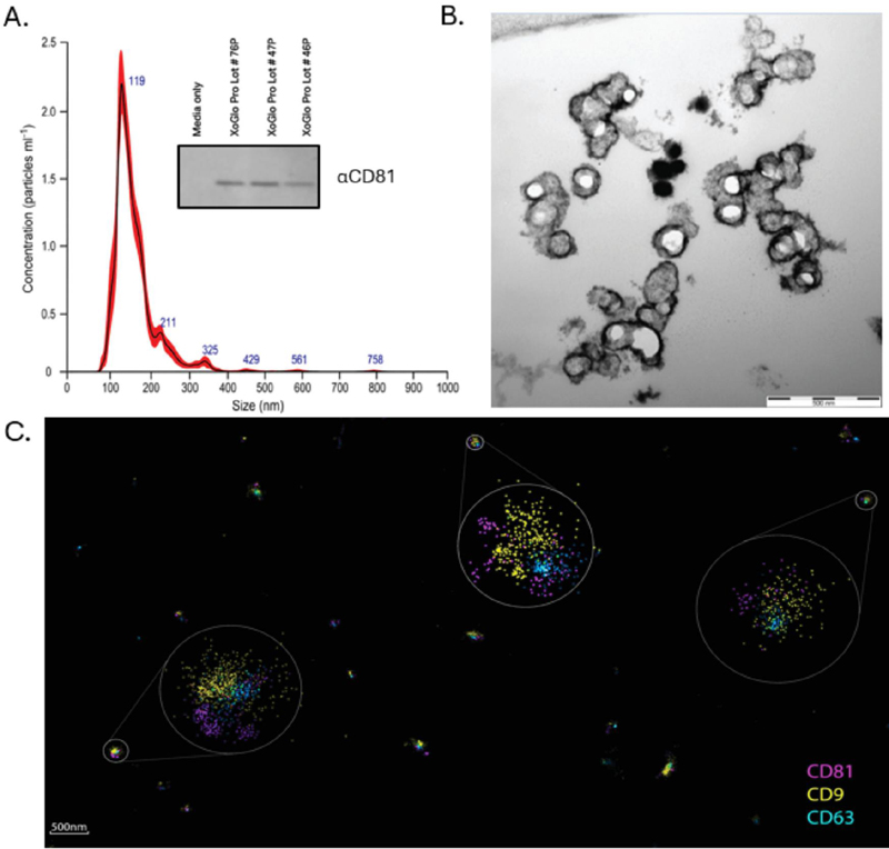
Figure 1 Multimodal characterization of placental MSC-derived exosomes (XoGlo Pro). (A) Nanoparticle tracking analysis (NanoSight) shows a predominant small-EV population (100–150 nm) with a minor larger fraction. Inset: Western blot detects the tetraspanins CD81 across independent production lots; conditioned media control is negative. (B) TEM reveals round, lipid bilayer vesicles with the characteristic cup-shaped morphology (scale bar as indicated). (C) dSTORM super-resolution imaging of individual vesicles immunolabeled for CD81 (magenta), CD9 (yellow), and CD63 (green) demonstrates tetraspanins co-localization and nanocluster organization on single exosomes (overview scale bar m; zooms as shown).
XoGlo® Pro Enhances Dermal Fibroblast Wound Closure
Human dermal fibroblast wound-closure assays were used to evaluate whether XoGlo® Pro altered cellular behaviors relevant to dermal repair. A standardized acellular gap was generated using chamber inserts, and fibroblast monolayers were treated with XoGlo® Pro or placebo/saline control conditions. Representative phase-contrast images demonstrated more rapid repopulation of the wound region in XoGlo® Pro-treated cultures compared with control cultures (Figure 2).
Quantitative image analysis showed a statistically significant reduction in residual gap area following XoGlo® Pro treatment relative to placebo/saline control conditions, with significance reported at p 0.01. Because mitomycin-C was not used, this assay measured the combined contribution of fibroblast migration and proliferation rather than migration alone. This design more closely reflects the coordinated cellular processes involved in dermal repair, where fibroblast motility, expansion, and matrix remodeling occur in parallel.
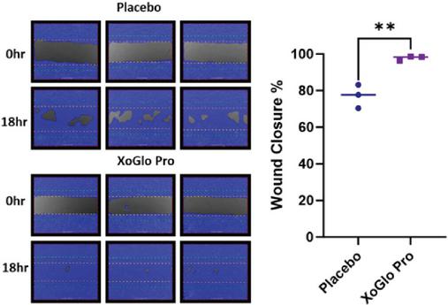
Figure 2 XoGlo® Pro treatment enhances wound closure. Human dermal fibroblasts were seeded in four chamber inserts. Inserts were removed and cells were then washed with PBS and then treated with either placebo or XoGlo® Pro mixed 1:1 with basal media. Cells were imaged daily for 7 days. FAST TRACK AI analyzed images to quantify gap closure after 18 h. Data is presented as a percentage of the time 0 h gap remaining at time 18 h. Data is presented as mean with standard deviation. **, value 0.01.
These findings indicate that XoGlo® Pro supports fibroblast-mediated closure of an in vitro wound model. In the context of potency assay development, the wound-closure assay provides a functional readout of regenerative activity relevant to dermal repair, but it should be interpreted alongside orthogonal assays evaluating viability, proliferation, phenotype, matrix deposition, and inflammatory modulation.
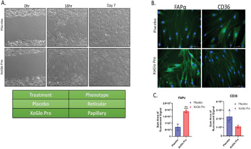
Figure 3 XoGlo® Pro treatment is associated with fibroblast morphology changes and selected phenotype-associated marker modulation. (Left) Scratch assay images show that placebo-treated fibroblasts primarily exhibit a reticular morphology, while XoGlo® Pro-treated fibroblasts exhibited an elongated papillary-like phenotype associated with enhanced non-fibrotic healing. (Right, top) Immunofluorescence staining demonstrates increased expression of fibroblast activation protein- (FAP, green) in XoGlo® Pro-treated cells compared to placebo, with nuclei counterstained in blue (DAPI). (Right, bottom) Quantification of fluorescence confirmed a significant increase in FAP expression in the XoGlo® Pro group (**, ), while CD36, a marker of fibrosis and inflammation was reduced. Statistical significance was evaluated using an unpaired two-tailed -test comparing XoGlo® Pro with placebo. Together, these findings are consistent with phenotype-associated modulation toward a papillary/pro-repair-associated fibroblast state.
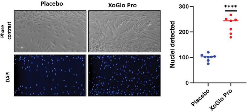
Figure 4 XoGlo® Pro enhances fibroblast density in the wound region. Phase-contrast microscopy (top panels) shows higher cell density and elongated morphology in XoGlo® Pro-treated fibroblasts compared with placebo. DAPI nuclear staining (bottom panels) demonstrates an increased number of nuclei in XoGlo® Pro-treated cultures. Quantification of cell counts (right) demonstrated a significant increase in fibroblast density following XoGlo® Pro treatment compared with placebo. Data are presented as individual replicate values. Statistical significance was evaluated using an unpaired two-tailed -test comparing XoGlo® Pro with placebo (****, ).
XoGlo® Pro Modulates Fibroblast Phenotype Toward a Pro-repair Profile
Fibroblast phenotype was assessed after prolonged culture in the wound-closure assay to determine whether XoGlo® Pro altered cellular morphology and marker expression in the repair region. Placebo-treated fibroblasts displayed a morphology consistent with a more reticular-like culture phenotype, whereas XoGlo® Pro-treated fibroblasts showed a more elongated and organized appearance consistent with a papillary/pro-repair-associated profile (Figure 3) [29, 30]. These morphological observations suggest phenotype-associated modulation under the assay conditions, rather than definitive lineage conversion [31]. Immunofluorescence analysis demonstrated increased fibroblast activation protein alpha signal in XoGlo® Pro-treated cultures compared with placebo controls. Quantitative analysis showed a significant increase in FAP-associated fluorescence intensity, reported at p 0.01 [32, 33]. In parallel, CD36 signal was reduced in XoGlo® Pro-treated cultures. Because CD36 has been associated with inflammatory and fibrotic fibroblast states in skin biology, its reduction is consistent with a shift away from a more pro-fibrotic or inflammatory phenotype [34]. These findings suggest that XoGlo® Pro may influence fibroblast state in a manner consistent with regenerative remodeling. Importantly, these data should be described as phenotype-associated modulation based on morphology and selected markers. Additional transcriptomic, proteomic, and functional matrix-remodeling assays would be needed to define fibroblast subtype identity more comprehensively.
XoGlo® Pro Increases Fibroblast Density and Cell Accumulation in the Wound-gap Region
Fibroblast accumulation and migration into the gap region was evaluated using phase-contrast imaging and DAPI-based nuclear quantification. XoGlo® Pro-treated cultures demonstrated visibly greater fibroblast density compared with placebo-treated controls (Figure 4). DAPI staining corroborated these observations by showing an increased number of nuclei in the XoGlo® Pro condition, consistent with enhanced cell accumulation within the assay period.
Quantitative cell-count analysis demonstrated a statistically significant increase in fibroblast number following XoGlo® Pro treatment, with significance reported at p 0.001. This accumulation response is relevant to dermal repair because fibroblast expansion and migration contributes to wound-bed repopulation, granulation tissue formation, extracellular matrix deposition, and tissue remodeling.
The data complement the wound-closure findings by supporting the interpretation that enhanced gap closure reflects both migration and proliferative expansion. In a translational potency framework, fibroblast proliferation provides a useful supportive readout of tissue repair activity, particularly when interpreted together with viability, phenotype modulation, and collagen deposition.
XoGlo® Pro Improves Fibroblast Viability Under Prolonged Culture Conditions
A live/dead assay was used to determine whether XoGlo® Pro supported fibroblast survival under prolonged culture conditions. Cells were stained with calcein AM to identify viable cells and Ethidium homodimer-1 to identify membrane-compromised cells. Representative fluorescence images showed a higher proportion of live cells in XoGlo® Pro-treated cultures compared with saline and comparator EV conditions shown in the figure (Figure 5). Quantification of the live/dead ratio demonstrated that XoGlo® Pro-treated fibroblasts maintained greater viability relative to the control and comparator conditions, with statistical significance reported at p 0.01. This survival-supportive effect suggests that XoGlo® Pro may contain bioactive cargo capable of supporting cellular stress resilience, metabolic activity, or anti-apoptotic signaling under the experimental conditions. Maintenance of fibroblast viability is important during normal tissue repair because fibroblasts contribute to wound-bed repopulation, extracellular matrix production, and subsequent tissue remodeling. However, increased fibroblast survival is not inherently anti-fibrotic. During physiological wound resolution, activated fibroblasts and myofibroblasts normally undergo phenotypic reversion, apoptosis, or clearance after sufficient matrix has been deposited. Persistent survival or activation of matrix-producing fibroblasts can instead promote excessive extracellular matrix accumulation, scar formation, and pathological fibrosis. Thus, the enhanced viability observed with XoGlo® Pro could theoretically represent an untoward pro-fibrotic effect if it were accompanied by sustained myofibroblast activation and excessive matrix deposition. The accompanying phenotype data in Figure 3 provide preliminary evidence that the enhanced fibroblast survival observed with XoGlo® Pro occurs in the context of a repair-associated rather than overtly fibrotic cellular state. XoGlo® Pro-treated fibroblasts exhibited an elongated morphology consistent with a more papillary/pro-repair-associated phenotype rather than the more reticular-like morphology observed in control cultures. This morphological shift was accompanied by increased FAP expression and reduced CD36 expression. FAP is associated with activated fibroblasts participating in tissue remodeling and repair, whereas reduced CD36 expression is directionally consistent with a shift away from fibroblast states associated with inflammatory and hypertrophic fibrotic remodeling. These findings are relevant to regenerative healing because maintenance of fibroblast viability is required for sustained repair, matrix production, and remodeling. However, the assay does not identify the specific molecular mechanism responsible for the survival effect. Future studies incorporating apoptosis markers, mitochondrial function, oxidative stress readouts, and cargo-depletion strategies would strengthen mechanistic interpretation.
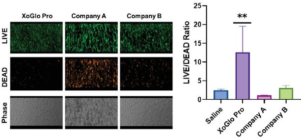
Figure 5 Live/dead cell viability assay of human dermal fibroblasts treated with exosome preparations. Cells were washed with PBS and incubated for 30 min in the dark with staining solution (1 l calcein AM, 20 l Ethidium homodimer-1 in 10 ml PBS; 750 l well-1). Live cells fluoresced green (calcein AM, GFP setting) and dead cells fluoresced orange/red (EthD-1, RFP setting). Representative fluorescence and phase-contrast images are shown (left). Quantification of the live/dead ratio (right) revealed that XoGlo Pro (Kimera Labs) significantly enhanced fibroblast viability compared to saline, Company A, and Company B controls (dose normalized to protein concentration). Data are presented as mean SD, . Statistical significance was evaluated by one-way ANOVA followed by Dunnett’s multiple-comparisons test, with XoGlo® Pro as the reference group (**, ).
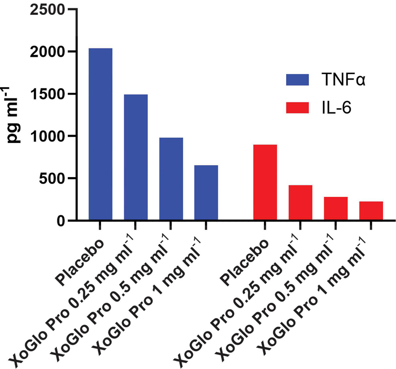
Figure 6 XoGlo® Pro attenuates LPS-induced TNF- and IL-6 secretion in macrophages. Rat alveolar macrophages (NR8383) were co-treated with LPS (100 ng ml-1) and XoGlo® Pro or with normal saline (placebo), diluted 1:1 with complete media. After 24 h conditioned media was collected and TNF- and IL-6 concentrations were quantified by ELISA. XoGlo® Pro reduced LPS-induced TNF- and IL-6 secretion in a concentration-associated manner. Data are expressed as pg ml-1.
XoGlo® Pro Attenuates Inflammatory Macrophage Cytokine Secretion
Macrophage inflammatory activation was evaluated following stimulation with LPS at a final concentration of 100 ng ml-1. LPS stimulation increased secretion of the pro-inflammatory cytokines TNF- and IL-6, consistent with activation of an inflammatory macrophage response (Figure 6). Co-treatment with XoGlo® Pro at 0.25, 0.5, and 1.0 mg ml-1 total protein concentration reduced secretion of both TNF- and IL-6 in a concentration-associated manner. Although XoGlo® Pro was independently characterized and quantified by NTA, the dosing series used in this macrophage assay was normalized according to total protein concentration in the final vial product, which was the experimental dose metric used when the assay was performed. NTA was used to characterize particle concentration and size distribution of the EV preparation, whereas protein concentration provided a practical normalization metric for comparing graded XoGlo® Pro treatment conditions in this functional assay. The findings support an immunomodulatory effect defined specifically by reduced TNF- and IL-6 secretion following LPS stimulation. This macrophage assay provides a mechanism-relevant functional readout for the immunomodulatory activity of XoGlo® Pro. In the context of tissue repair and healthy longevity, attenuation of excessive inflammatory cytokine signaling may be relevant to chronic inflammation, impaired wound healing, immune dysregulation, and inflammatory tissue injury. Additional studies incorporating broader cytokine panels, NF-B and MAPK pathway activation, transcriptomic profiling, phagocytic function, and other macrophage functional endpoints would further define the underlying mechanism of action.
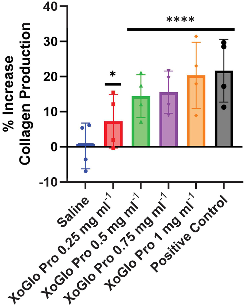
Figure 7 XoGlo® Pro increases collagen-associated deposition in human dermal fibroblast cultures. Human dermal fibroblasts were treated for 48 h with XoGlo® Pro at the indicated concentrations in a 1:1 mixture with complete culture medium. Collagen-associated deposition was assessed using a Sirius Red dye-binding assay and normalized to the saline vehicle control. TGF-1 (5 ng ml-1) served as the positive control. Data are presented as mean SD with individual replicate values overlaid. Statistical significance was evaluated by one-way ANOVA followed by Dunnett’s multiple-comparisons test, with the saline vehicle control as the reference group. *, and ****, versus saline control.
XoGlo® Pro Enhances Collagen Deposition by Human Dermal Fibroblasts
Collagen deposition was measured using Sirius Red dye-binding in human dermal fibroblast cultures treated with increasing concentrations of XoGlo® Pro. Saline served as the negative control, untreated cells provided an additional baseline reference, and TGF-1 was included as a positive collagen-inducing control (Figure 7). XoGlo® Pro increased collagen deposition relative to saline control conditions, with the highest tested concentration approaching the effect observed with TGF-1 in the assay shown.
The concentration-associated increase in Sirius Red signal indicates that XoGlo® Pro supports extracellular matrix production by dermal fibroblasts under the experimental conditions. This matrix-supportive activity is biologically relevant to wound repair, dermal remodeling, and regenerative tissue responses. When interpreted with the fibroblast phenotype data, increased collagen deposition suggests support for matrix remodeling rather than a simple pro-fibrotic response. The highest tested XoGlo® Pro concentration produced a Sirius Red signal within the range of the TGF-1 positive-control response in this assay. Because Sirius Red quantifies collagen-associated dye binding but does not define matrix quality, collagen subtype, or fibrotic phenotype, these findings require interpretation alongside fibrosis-associated markers and matrix-architecture studies [35].
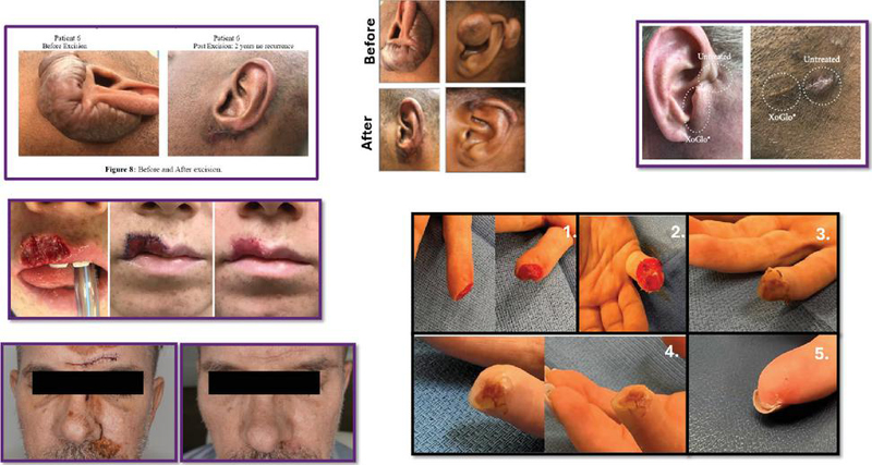
Figure 8 Representative before-and-after clinical case images following use of XoGlo® Pro in dermatologic and wound-repair settings. De-identified images supplied by clinical investigators or treating clinicians illustrate observed tissue-repair outcomes across multiple real-world case examples, including post-excision auricular healing, localized skin/ear treatment comparisons, traumatic lip and facial wounds, and a distal fingertip injury followed over serial healing time points. Images were obtained either in IRB-approved studies or in clinical practice under informed consent, as applicable. These photographs are presented as observational case-study materials to document clinical experience and should not be interpreted as controlled efficacy evidence. Interpretation is limited by differences in indication, treatment setting, timing of image capture, photography conditions, wound severity, concomitant care, and absence of standardized blinded assessment unless otherwise specified.
Observational Clinical Case Images Provide Real-World Tissue-Repair Context
De-identified before-and-after clinical images were included to provide real-world observational context for dermatologic and wound-repair use of XoGlo® Pro (Figure 8). Clinical images were provided by treating clinicians, clinical investigators, or external clinical contributors under applicable informed consent or IRB-approved protocols. The cases include examples of dermatologic repair, post-procedural healing, traumatic injury, and serial wound follow-up.
Across the representative images, visible changes in tissue appearance were observed over time, including apparent wound closure, improved epithelial coverage, reduced visible injury burden, and progressive tissue remodeling in selected cases. These observations are consistent with the in vitro findings showing fibroblast wound closure, viability support, matrix deposition, and immunomodulatory activity. However, the images are observational and were not generated under a standardized controlled imaging protocol unless otherwise specified.
These clinical case materials should not be interpreted as controlled efficacy evidence. Their interpretation is limited by variable indications, wound severity, treatment conditions, concomitant care, follow-up timing, camera angle, lighting, distance, image quality, and the absence of blind independent assessment. Their value is translational and contextual: they illustrate ethically documented clinical observations that may inform future controlled study design, endpoint selection, and patient-access pathways.
IRB-approved Proof-of-Concept Longevity Study Shows Exploratory Functional and Cognitive Signals
An IRB-approved proof-of-concept clinical study was used to evaluate XoGlo® Pro in a healthy longevity-related context (Figure 9). The study assessed safety and exploratory changes in patient-reported function, fatigue, mood, immune-related measures, and cognitive performance over the follow-up period. The figure presents individual participant-level changes from baseline to follow-up, with group means shown for selected domains.
XoGlo® Pro administration was not associated with serious adverse events in the study population described in the draft. Directional improvements were observed across several exploratory domains, including patient-reported functional status, fatigue, mood-related measures, and cognitive performance. These findings are consistent with the broader translational hypothesis that placental MSC-derived EVs may influence biological pathways relevant to immune modulation, tissue repair signaling, functional resilience, and cognitive performance.
These clinical findings should be interpreted as exploratory proof-of-concept observations rather than definitive efficacy evidence. The study was not designed or powered to establish clinical effectiveness, and the absence of a large randomized controlled comparison limits causal inference. Nevertheless, the combination of safety observations and directional functional-cognitive signals provides a rationale for future controlled studies evaluating XoGlo® Pro in healthy longevity populations using prespecified endpoints, blinded assessment, appropriate comparators, longer follow-up, and biomarker-linked outcome analysis.
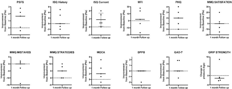
Figure 9 Exploratory functional, cognitive, and patient-reported changes from baseline to 1-month follow-up. Individual participant changes from baseline to 1-month follow-up are shown across cognitive, mood, immune-related, and functional measures. Each point represents a single participant, with group means indicated. Individual participant-level exploratory measures showed directional changes from baseline to 1-month follow-up.
Integrated Translational Interpretation
Across the analytical, in vitro, observational, and proof-of-concept clinical components of this study, XoGlo® Pro demonstrated a coherent translational profile consistent with a placental MSC-derived EV regenerative platform. Multimodal characterization supported small-EV enrichment, EV-associated marker detection, vesicle morphology, and lot characterization. Functional assays demonstrated activities relevant to regenerative healing and immune modulation, including enhanced fibroblast wound closure, proliferation, viability, phenotype-associated modulation, collagen deposition, and attenuation of inflammatory macrophage cytokine secretion.
The observational clinical case images and IRB-approved proof-of-concept longevity study extend these findings into ethically documented human-use contexts while remaining appropriately limited in interpretation. Together, these data support further clinical investigation of XoGlo® Pro as a placental MSC-EV platform for immune modulation, tissue repair, healthy longevity, functional resilience, and cognitive performance. Future studies should prioritize controlled trial design, standardized imaging, validated functional and cognitive endpoints, biomarker-linked mechanisms, lot-to-lot potency correlation, and long-term safety monitoring.
Discussion
This original translational study evaluated XoGlo® Pro, a placental mesenchymal stromal/stem cell-derived small EV-enriched preparation, using an integrated framework that combined analytical characterization, mechanism-relevant in vitro assays, observational clinical case materials, and an IRB-approved proof-of-concept healthy longevity study. The analytical data supported a small-EV-enriched product profile, including nanoscale particle distribution by nanoparticle tracking analysis, vesicle morphology by transmission electron microscopy, EV-associated marker detection by immunoblotting and dSTORM (Figure 1). This type of orthogonal characterization is consistent with current extracellular vesicle reporting standards, which emphasize that EV preparations should be defined by complementary measurements rather than a single marker, particle count, or imaging modality [36, 37]. Importantly, these data support product identity, lot characterization, and mechanism-supportive cargo assessment, but they do not independently establish biological potency.
The in vitro findings demonstrated biological activities relevant to regenerative healing and immune modulation. In human dermal fibroblasts, XoGlo® Pro enhanced gap closure in a scratch/gap assay (Figure 2), supported phenotype-associated modulation toward a papillary/pro-repair profile (Figure 3), increased fibroblast proliferation (Figure 4), improved viability under prolonged culture conditions (Figure 5), and increased collagen deposition (Figure 7). In LPS-stimulated macrophages, XoGlo® Pro reduced secretion of inflammatory cytokines, including TNF- and IL-6 (Figure 6). Together, these findings suggest that XoGlo® Pro may act through multiple repair-relevant and immunomodulatory mechanisms rather than through a single isolated pathway. This interpretation is consistent with the broader MSC-EV literature, which supports the concept that MSC-derived EVs transfer complex regulatory cargo, including miRNAs, mRNAs, proteins, lipids, and other signaling molecules, to recipient cells and thereby influence inflammation, angiogenesis, extracellular matrix remodeling, and tissue repair [1, 4, 18, 38].
The fibroblast assay panel provides a biologically plausible basis for interpreting the dermal repair findings. Cutaneous repair requires coordinated fibroblast migration, proliferation, survival, extracellular matrix deposition, and remodeling. MSC-EVs have been reported in preclinical wound models to promote wound closure, increase fibroblast and keratinocyte migration/proliferation, support angiogenesis, and modulate inflammatory responses during the transition from inflammatory to proliferative phases of repair [14, 38]. In this study, XoGlo® Pro enhanced gap closure in the wound-closure assay (Figure 2), increased fibroblast density and nuclear counts (Figure 4), improved live/dead ratios under prolonged culture conditions (Figure 5), and increased collagen deposition by Sirius Red staining (Figure 7). Because mitomycin-C was not used, the wound-closure assay reflects combined migration and proliferation rather than migration alone. This is appropriate for a repair-focused model because wound closure in vivo depends on integrated cellular movement, expansion, matrix deposition, and remodeling.
The fibroblast phenotype data suggests that XoGlo® Pro may influence dermal fibroblast state. Treated cultures displayed morphology consistent with a papillary/pro-repair-associated profile, increased FAP signal, and reduced CD36 signal compared with controls (Figure 3). These observations are best interpreted as phenotype-associated modulation rather than definitive fibroblast lineage conversion. Fibroblast populations in skin are heterogeneous and context-dependent, and individual markers may reflect activation state, metabolic state, inflammatory exposure, or matrix-remodeling activity rather than stable lineage identity. Similarly, increased Sirius Red staining supports matrix-associated activity (Figure 7), but collagen quantity alone does not distinguish regenerative matrix formation from fibrotic remodeling. Future studies should evaluate collagen I/III ratio, matrix organization, -SMA expression, myofibroblast contractility, fibrotic transcriptional programs, and single-cell or spatial transcriptomic features to better define whether XoGlo® Pro supports regenerative remodeling rather than scar-promoting fibrosis.
The macrophage data provide complementary evidence of immunomodulatory activity. LPS stimulation induced secretion of TNF- and IL-6, while XoGlo® Pro reduced these inflammatory cytokines under the assay conditions tested (Figure 6). This supports attenuation of selected inflammatory responses and is consistent with the broader literature showing that MSCs and MSC-EVs can influence macrophage inflammatory programs, including suppression of pro-inflammatory cytokines and promotion of resolution-associated phenotypes in multiple tissue-injury models [39–41].
The characterization and functional assay strategy used here reflects a central challenge in EV biologic development. NTA, TEM, immunoblotting, dSTORM, and small RNA profiling (data not shown) each contribute important information about particle size, morphology, purity-related attributes, EV-associated markers, molecular heterogeneity, and cargo composition (Figure 1). However, particle concentration, tetraspanin detection, and miRNA cargo do not independently define potency. Current EV-development literature emphasizes that therapeutic EV products require mechanism-linked functional assays, reference standards, lot comparability, dose normalization, and stability-indicating readouts [1, 4, 18, 36, 37]. In this study, fibroblast wound closure (Figure 2), phenotype-associated marker expression (Figure 3), proliferation (Figure 4), viability (Figure 5), macrophage cytokine suppression (Figure 6), and collagen deposition (Figure 7) provide a candidate functional potency framework for dermal repair and immune modulation. Further assay qualification will be needed to establish precision, linearity, sensitivity, specificity, robustness, reference-standard performance, lot-discriminating capacity, and stability-indicating relevance.
Placental MSC-derived EVs are of particular translational interest because placental tissue is a perinatal source associated with fetal-maternal immune tolerance, growth-factor signaling, vascular remodeling, and tissue development. These biological attributes provide a strong rationale for studying placental MSC-EVs in tissue repair and immune modulation. However, tissue source should be treated as a biological rationale rather than a substitute for product-specific evidence. EV cargo and function can vary with donor, tissue source, cell-culture conditions, passage, oxygen tension, inflammatory priming, isolation method, purification strategy, storage, and dose metric [1, 4, 18, 37]. Therefore, XoGlo® Pro should be evaluated as a defined biologic product whose translational value depends on reproducible manufacturing, analytical profile, cargo composition, functional potency, safety attributes, and clinical evidence.
The healthy-longevity component of this study should be interpreted in the context of emerging preclinical evidence linking MSC-EVs to senescence biology and functional resilience. Aging is associated with impaired tissue repair, chronic low-grade inflammation, stem-cell dysfunction, cellular senescence, mitochondrial dysfunction, and altered intercellular communication [10, 42]. In mouse models, MSC-derived EVs have been reported to reduce senescence markers, rescue aged stem-cell and fibroblast function, and improve healthspan-related outcomes [43]. These findings provide mechanistic rationale for studying MSC-EVs in healthy-longevity contexts, but they do not establish clinical efficacy in humans. In the present study, XoGlo® Pro administration was not associated with serious adverse events in the proof-of-concept cohort and was accompanied by directional improvements in patient-reported function, fatigue, mood, and cognitive performance (Figure 9). These observations are hypothesis-generating and should be tested in controlled studies with prespecified functional, cognitive, immune, inflammatory, and biomarker-linked endpoints.
The broader relevance of MSC-EVs to musculoskeletal decline and neuroinflammation also supports future indication-specific development. Preclinical studies suggest that MSC-EVs can influence skeletal muscle regeneration, satellite-cell activity, myogenic differentiation, inflammation, and muscle atrophy-related pathways [44–48]. Other studies in traumatic brain injury models have reported that MSC-derived exosomes can reduce neuroinflammation, suppress microglial/macrophage activation, reduce neuronal apoptosis, and improve functional recovery [49–51]. These findings are relevant to the manuscript’s interest in functional resilience and cognitive outcomes, but they should be used only as mechanistic context. The present study did not directly assess biodistribution, EV uptake in neural or muscle tissues, neuroinflammatory biomarkers, muscle regeneration endpoints, or causal links between EV cargo and cognitive-functional outcomes. Future studies should include indication-specific models and biomarkers before extending claims beyond dermal repair and immune modulation.
The human-context data extend the translational relevance of the analytical and in vitro findings while remaining exploratory. De-identified before-and-after images supplied by clinicians, or investigators from IRB-approved studies or informed-consent clinical practice illustrate tissue-repair observations across dermatologic and wound-repair settings (Figure 8). These images are useful for documenting real-world clinical experience and informing future endpoint selection, imaging procedures, and controlled study design. However, they are not controlled efficacy evidence. Interpretation is limited by variability in wound type, severity, anatomic location, treatment timing, concomitant care, camera angle, lighting, distance, image quality, and absence of blinded standardized assessment unless otherwise specified. Future dermal and wound-repair studies should incorporate standardized photography, calibrated wound measurement, blinded adjudication, validated wound-severity scales, patient-reported outcomes, and predefined healing endpoints.
Regulatory translation of EV-based biologics requires disciplined chemistry, manufacturing, and controls development. Key considerations include donor eligibility, tissue traceability, cell banking, raw material control, aseptic processing, sterility, endotoxin, mycoplasma testing, residual process reagent assessment, formulation, storage, stability, lot comparability, and dose normalization. EV-specific challenges include product heterogeneity, limited interpretability of single-marker identity assays, cargo variability, particle-to-protein ratio interpretation, uncertainty around optimal dose metrics, and incomplete mechanism-of-action definition. Recent analyses of MSC-EV clinical trials highlight substantial variability in EV characterization, dose units, route of administration, and outcome reporting, emphasizing the need for harmonized dosing frameworks, potency assays, and clinical protocols [52]. For XoGlo® Pro, the integration of orthogonal characterization (Figure 1) with mechanism-linked functional assays (Figures 2–7) provides a translational foundation, but further development should include reference standards, comparability criteria, qualified potency assays, stability-indicating functional readouts, dose-response studies, and correlation of in vitro activity with clinical biomarkers and outcomes.
This study has several limitations. The in vitro assays model selected aspects of dermal repair and immune modulation but cannot reproduce the full complexity of human tissue injury, vascularization, innervation, immune-cell diversity, wound microbiology, systemic inflammation, or aging biology. Mechanistic interpretation is limited by selected marker panels and functional readouts, including fibroblast phenotype markers (Figure 3), macrophage cytokines (Figure 6), and collagen deposition (Figure 7). Additional studies are needed to confirm EV uptake, cargo transfer, target engagement, downstream signaling, fibroblast subtype identity, macrophage state, matrix organization, angiogenesis, senescence modulation, biodistribution, pharmacodynamics, and durability of response. The observational clinical images are subject to selection bias and non-standardized imaging conditions (Figure 8). The proof-of-concept longevity study involved a limited sample size, short follow-up, and was not powered to establish efficacy; placebo effects, regression to the mean, natural variability, and background care must be considered (Figure 9).
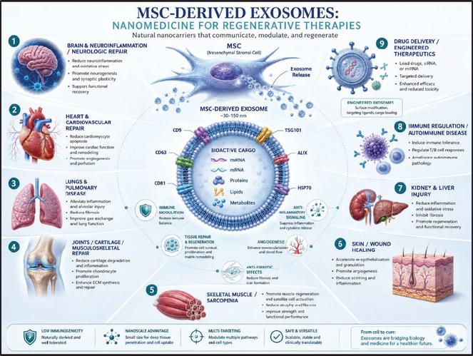
Figure 10 Therapeutic rationale for MSC-derived exosomes in regenerative medicine. MSC-derived exosomes are nanoscale extracellular vesicles released by mesenchymal stromal/stem cells that carry bioactive cargo, including miRNA, mRNA, proteins, lipids, and metabolites. Through intercellular communication, these vesicles may modulate inflammation, support tissue repair, promote angiogenesis, influence fibrosis, and regulate cellular stress responses. The diagram summarizes potential regenerative and translational application areas, including neuroinflammation and neurologic repair, cardiovascular repair, pulmonary disease, musculoskeletal injury, sarcopenia, wound healing, kidney and liver injury, immune regulation, and engineered drug delivery. These proposed applications remain indication- and product-specific and require rigorous characterization, potency testing, non-clinical validation, and controlled clinical studies.
Future work should prioritize controlled clinical trials in defined indications such as dermal repair, wound healing, functional decline, and healthy longevity. These studies should include standardized clinical photography informed by the limitations of the current case-image dataset (Figure 8), blinded wound assessment, validated cognitive and functional endpoints, patient-reported outcomes, inflammatory and immune biomarker panels, dose-response analysis, and long-term safety monitoring. Mechanistic studies should incorporate transcriptomics, proteomics, miRNA cargo-function mapping based on the characterization strategy shown in Figure 1, EV uptake assays, macrophage phenotype markers, fibroblast subtype profiling, collagen subtype analysis, matrix architecture, angiogenesis assays, mitochondrial stress models, senescence/inflammaging systems, and cargo-depletion or pathway-inhibition experiments. Linking lot-level potency data to clinical biomarker and outcome measures will be essential for determining which assays are most predictive of biological activity in humans.
Overall, the combined analytical, in vitro, observational, and proof-of-concept clinical findings support further clinical investigation of XoGlo® Pro as a placental MSC-derived small EV-enriched regenerative platform. The data support small-EV enrichment, EV-associated marker detection, fibroblast repair-associated activity, matrix-supportive effects, and attenuation of selected macrophage inflammatory cytokine responses (Figures 1–7). Human-context observations provide additional translational relevance but remain exploratory and non-definitive (Figures 8 and 9). The therapeutic rationale diagram provides a conceptual framework for broader MSC-EV regenerative applications while emphasizing the need for indication-specific validation (Figure 10). Future controlled studies with qualified potency assays, validated endpoints, biomarker-linked mechanisms, standardized imaging, and long-term safety monitoring are warranted to define the clinical potential of XoGlo® Pro in immune modulation, tissue repair, healthy longevity, functional resilience, and cognitive performance.
Ethics Statement
The human proof-of-concept longevity study was reviewed and approved by the GARM International Foundation Institutional Review Board (IRB) under study approval GIFIRB202506191 (IRB Unique Identifier: RESET-REV), with approval granted on November 26, 2025. The approved protocol was titled “Phase IIa Trial to Evaluate the Safety and Efficacy of Placental Mesenchymal Stem Cell-Derived Extracellular Vesicles Combined with Low-Intensity Focused Ultrasound Among Patients with Sarcopenia.” The proof-of-concept cohort of patients reported in this paper evaluated intravenous administration of XoGlo® Pro as an exploratory intervention in the context of healthy longevity and age-related functional decline as part of a larger study still ongoing. All participants provided written informed consent prior to participation. The study was conducted in accordance with the IRB-approved protocol, applicable ethical and regulatory requirements, and internationally recognized principles for the protection of human research participants. Where applicable, clinical activities were additionally conducted under relevant local regulatory authorization. Clinical operations in The Bahamas were authorized through the applicable national regulatory framework, including oversight by the Bahamas Ministry of Health and Wellness and the National Longevity and Regenerative Therapies Board (LARTA). These local authorizations were distinct from the independent IRB review and approval of the research protocol.
Conflict of Interest Statement
Dillon Betancourt may hold employment with Kimera Labs, Inc., the developer, and manufacturer of XoGlo® Pro. Duncan Ross III is the founder and Chief Scientific Officer of Kimera Labs, Inc. The authors may hold employment, equity, intellectual property, consulting, or financial interests related to Kimera Labs, Inc., and/or XoGlo® Pro. The studies described in this paper include data generated using XoGlo® Pro, a Kimera Labs product. These relationships represent potential conflicts of interest and are disclosed for transparency. The authors state that the manuscript presents analytical, in vitro, observational, and proof-of-concept clinical findings with appropriate limitations and does not claim established clinical efficacy.
References
[1] M. Gimona et al., “Critical considerations for the development of potency tests for therapeutic applications of mesenchymal stromal cell-derived small extracellular vesicles,” Cytotherapy, vol. 23, no. 5, pp. 373–380, 2021.
[2] C. Raggi and A. C. Berardi, “Mesenchymal stem cells, aging and regenerative medicine,” Muscles Ligaments Tendons J., vol. 2, no. 3, pp. 239–242, 2012.
[3] M. D. Hade, C. N. Suire, and Z. Suo, “Mesenchymal stem cell-derived exosomes: Applications in regenerative medicine,” Cells, vol. 10, no. 8, 2021.
[4] T. Lener et al., “Applying extracellular vesicles based therapeutics in clinical trials – an ISEV position paper,” J. Extracell. Vesicles, vol. 4, p. 30087, 2015.
[5] A. T. Reiner et al., “Concise review: Developing best-practice models for the therapeutic use of extracellular vesicles,” Stem Cells Transl. Med., vol. 6, no. 8, pp. 1730–1739, 2017.
[6] B. Claridge et al., “Development of extracellular vesicle therapeutics: Challenges, considerations, and opportunities,” Front. Cell Dev. Biol., vol. 9, p. 734720, 2021.
[7] M. Gimona et al., “Manufacturing of human extracellular vesicle-based therapeutics for clinical use,” Int. J. Mol. Sci., vol. 18, no. 6, 2017.
[8] E. M. Crimmins, “Lifespan and healthspan: Past, present, and promise,” Gerontologist, vol. 55, no. 6, pp. 901–911, 2015.
[9] J. J. Baechle et al., “Chronic inflammation and the hallmarks of aging,” Mol. Metab., vol. 74, p. 101755, 2023.
[10] C. López-Otín et al., “Hallmarks of aging: An expanding universe,” Cell, vol. 186, no. 2, pp. 243–278, 2023.
[11] Y. Zhu et al., “Application of mesenchymal stem cell therapy for aging frailty: From mechanisms to therapeutics,” Theranostics, vol. 11, no. 12, pp. 5675–5685, 2021.
[12] Z. Zhao et al., “The role of mesenchymal stem cell-derived exosome in epigenetic modifications in inflammatory diseases,” Front. Immunol., vol. 14, p. 1166536, 2023.
[13] P. Zhao et al., “Exosomes derived from bone marrow mesenchymal stem cells improve osteoporosis through promoting osteoblast proliferation via MAPK pathway,” Eur. Rev. Med. Pharmacol. Sci., vol. 22, no. 12, pp. 3962–3970, 2018.
[14] Q. L. Zeng and D. W. Liu, “Mesenchymal stem cell-derived exosomes: An emerging therapeutic strategy for normal and chronic wound healing,” World J. Clin. Cases, vol. 9, no. 22, pp. 6218–6233, 2021.
[15] A. Uccelli, L. Moretta, and V. Pistoia, “Mesenchymal stem cells in health and disease,” Nat. Rev. Immunol., vol. 8, no. 9, pp. 726–736, 2008.
[16] Y. Shahsavandi et al., “miRNAs from mesenchymal-stem-cell-derived extracellular vesicles: Emerging players in regenerative medicine and disease therapy,” Mol. Ther. Nucleic Acids, vol. 36, no. 4, 2025.
[17] S. Maacha et al., “Paracrine mechanisms of mesenchymal stromal cells in angiogenesis,” Stem Cells Int., vol. 2020, p. 4356359, 2020.
[18] K. W. Witwer et al., “Defining mesenchymal stromal cell (MSC)-derived small extracellular vesicles for therapeutic applications,” J. Extracell. Vesicles, vol. 8, no. 1, p. 1609206, 2019.
[19] M. R. Mohammadi et al., “Isolation and characterization of microvesicles from mesenchymal stem cells,” Methods, vol. 177, pp. 50–57, 2020.
[20] K. Yin, S. Wang, and R. C. Zhao, “Exosomes from mesenchymal stem/stromal cells: A new therapeutic paradigm,” Biomark. Res., vol. 7, p. 8, 2019.
[21] F. Tan et al., “Clinical applications of stem cell-derived exosomes,” Signal Transduct. Target. Ther., vol. 9, no. 1, p. 17, 2024.
[22] Y. S. Chen et al., “Exosomes in clinical trial and their production in compliance with good manufacturing practice,” Ci Ji Yi Xue Za Zhi, vol. 32, no. 2, pp. 113–120, 2020.
[23] L. Wang et al., “Characterization of placenta-derived mesenchymal stem cells cultured in autologous human cord blood serum,” Mol. Med. Rep., vol. 6, no. 4, pp. 760–766, 2012.
[24] M. Wu et al., “Comparison of the biological characteristics of mesenchymal stem cells derived from the human placenta and umbilical cord,” Sci. Rep., vol. 8, no. 1, 2018.
[25] M. Komaki et al., “Exosomes of human placenta-derived mesenchymal stem cells stimulate angiogenesis,” Stem Cell Res. Ther., vol. 8, no. 1, p. 219, 2017.
[26] A. Papait et al., “Mesenchymal stromal cells from fetal and maternal placenta possess key similarities and differences: Potential implications for their applications in regenerative medicine,” Cells, vol. 9, no. 1, 2020.
[27] Y. Zhu et al., “Placental mesenchymal stem cells of fetal and maternal origins demonstrate different therapeutic potentials,” Stem Cell Res. Ther., vol. 5, no. 2, p. 48, 2014.
[28] R. A. Pelekanos et al., “Isolation and expansion of mesenchymal stem/stromal cells derived from human placenta tissue,” J. Vis. Exp., no. 112, 2016.
[29] D. G. Janson et al., “Different gene expression patterns in human papillary and reticular fibroblasts,” J. Invest. Dermatol., vol. 132, no. 11, pp. 2565–2572, 2012.
[30] A. Korosec et al., “Lineage identity and location within the dermis determine the function of papillary and reticular fibroblasts in human skin,” J. Invest. Dermatol., vol. 139, no. 2, pp. 342–351, 2019.
[31] D. Janson et al., “Papillary fibroblasts differentiate into reticular fibroblasts after prolonged in vitro culture,” Exp. Dermatol., vol. 22, no. 1, pp. 48–53, 2013.
[32] J. E. Park et al., “Fibroblast activation protein, a dual specificity serine protease expressed in reactive human tumor stromal fibroblasts,” J. Biol. Chem., vol. 274, no. 51, pp. 36505–36512, 1999.
[33] J. Roman, “Fibroblasts – warriors at the intersection of wound healing and disrepair,” Biomolecules, vol. 13, no. 6, 2023.
[34] M. F. Griffin et al., “JUN promotes hypertrophic skin scarring via CD36 in preclinical in vitro and in vivo models,” Sci. Transl. Med., vol. 13, no. 609, p. eabb3312, 2021.
[35] P. Lin, G. Zhang, and H. Li, “The role of extracellular matrix in wound healing,” Dermatol. Surg., vol. 49, no. 5S, pp. S41–S48, 2023.
[36] C. Théry et al., “Minimal information for studies of extracellular vesicles 2018 (MISEV2018): A position statement of the International Society for Extracellular Vesicles and update of the MISEV2014 guidelines,” J. Extracell. Vesicles, vol. 7, no. 1, p. 1535750, 2018.
[37] J. A. Welsh et al., “Minimal information for studies of extracellular vesicles (MISEV2023): From basic to advanced approaches,” J. Extracell. Vesicles, vol. 13, no. 2, p. e12404, 2024.
[38] C. McLaughlin et al., “Mesenchymal stem cell-derived extracellular vesicles for therapeutic use and in bioengineering applications,” Cells, vol. 11, no. 21, 2022.
[39] S. S. Kuppa et al., “Role of mesenchymal stem cells and their paracrine mediators in macrophage polarization: An approach to reduce inflammation in osteoarthritis,” Int. J. Mol. Sci., vol. 23, no. 21, p. 13016, 2022.
[40] Y. Shi et al., “Exosomes derived from bone marrow stromal cells (BMSCs) enhance tendon-bone healing by regulating macrophage polarization,” Med. Sci. Monit., vol. 26, p. e923328, 2020.
[41] X. He et al., “MSC-derived exosome promotes M2 polarization and enhances cutaneous wound healing,” Stem Cells Int., vol. 2019, p. 7132708, 2019.
[42] J. Boulestreau et al., “Mesenchymal stem cell derived extracellular vesicles in aging,” Front. Cell Dev. Biol., vol. 8, p. 107, 2020.
[43] A. Dorronsoro et al., “Mesenchymal stem cell-derived extracellular vesicles reduce senescence and extend health span in mouse models of aging,” Aging Cell, vol. 20, no. 4, p. e13337, 2021.
[44] K. A. Cho et al., “Mesenchymal stem cell-derived exosomes protect muscle loss by miR-145-5p activity targeting activin A receptors,” Cells, vol. 10, no. 8, 2021.
[45] S. E. Byun et al., “Skeletal muscle regeneration by the exosomes of adipose tissue-derived mesenchymal stem cells,” Curr. Issues Mol. Biol., vol. 43, no. 3, pp. 1473–1488, 2021.
[46] E. Mahindran et al., “Mesenchymal stem cell-derived extracellular vesicles: Hype or hope for skeletal muscle anti-frailty,” Int. J. Mol. Sci., vol. 24, no. 9, 2023.
[47] C. Porcu, G. Dobrowolny, and B. M. Scicchitano, “Exploring the role of extracellular vesicles in skeletal muscle regeneration,” Int. J. Mol. Sci., vol. 25, no. 11, 2024.
[48] K. Wang et al., “Nanotopographical cues tune the therapeutic potential of extracellular vesicles for the treatment of aged skeletal muscle injuries,” ACS Nano, vol. 17, no. 20, pp. 19640–19651, 2023.
[49] H. Ni et al., “Exosomes derived from bone mesenchymal stem cells ameliorate early inflammatory responses following traumatic brain injury,” Front. Neurosci., vol. 13, no. 14, 2019.
[50] Y. Chen et al., “MSC-derived exosomes promote recovery from traumatic brain injury via microglia/macrophages in rat,” Aging (Albany NY), vol. 12, no. 18, pp. 18274–18296, 2020.
[51] L. Wen et al., “Exosomes derived from bone marrow mesenchymal stem cells inhibit neuroinflammation after traumatic brain injury,” Neural Regen. Res., vol. 17, no. 12, pp. 2717–2724, 2022.
[52] Y. Wang et al., “Trends in mesenchymal stem cell-derived extracellular vesicles clinical trials 2014–2024: Is efficacy optimal in a narrow dose range?” Front. Med., vol. 12, p. 1625787, 2025.
International Journal of Translational Science, 2026, 123–158
doi: 10.13052/ijts2246-8765.2026.006
© 2026 River Publishers