Deep Learning Technique Based State-Of-The-Art in Skin Cancer Detection: A Review
CH. Srilakshmi1,*, E. Laxmi Lydia2 and N. Ramakrishnaiah3
1Department of Information Technology, Malla Reddy Engineering College for Women, Telangana, India
2Department of Computer Science and Engineering, Vignan’s Institute of Information Technology, Visakhapatnam, India
3Department of CSE, University College of Engineering, JNTU Kakinada, Andhra Pradesh, India
E-mail: srilaks_ch99@outlook.com; chsrilu.45@gmail.com; nrkrishna27@jntucek.ac.in
*Corresponding Author
Received 14 December 2022; Accepted 03 July 2023; Publication 13 October 2023
Abstract
Research of skin cancer images through visual survey and manual evaluation to investigate skin threatening development has always been abnormal. This manual evaluation of skin injuries to recognize melanoma is monotonous as well as somewhat long. With movement in advancement and fast development in computational resources, different AI techniques and significant learning methods have emerged for assessment of clinical pictures most especially the skin lesion images. In late years, AI arising as an innovation equipped for tackling issues connected with horticulture, medical services, business, and soon. To diminish the endanger to human existence we can embrace AI calculations in the medical care area and can foresee the deadliest skin illnesses like dangerous melanoma in beginning phases. The point of the research is to give bits of knowledge about various classifications of skin lesions and strategies executed to arrange and foresee skin diseases and the job of dermatologists while fostering the models, at last gives a general rundown of existing work.
Keywords: Skin lesion, melanoma, machine learning algorithms, malignant, skin cancers.
1 Introduction
Disease is an extreme life risk to human life. It could at times make inescapable downfall the human. Different kinds of harmful development could exist in human body as well as skin sickness is maybe of fastest creating threatening development that can cause passing. The greatest effective technique to stop the transmission of skin conditions in any situation is through hand washing (hand hygiene). To thoroughly rinse your hands: Use cleanser after washing the wrists with safe water. It is instigated by specific factors responsive qualities, defilements, contaminations, genuine work, regular change, receptiveness to bright light, and so on [1]. Substances such polychlorinated biphenyls (PCBs) & dioxin, that are present at a certain polluted site, often cause individuals to be concerned. Daily-use items like cleaning chemicals, prescriptions as well as over medications, liquor, petroleum, insecticides, heating oil, and perfumes can all be dangerous. Moreover, unusual swellings of human body are similarly a justification behind skin harmful development, most unremitting sorts of skin harmful development [2]. The world wellbeing association (WHO) predicts that one in every three illnesses examined is skin dangerous development. The amount of people who are examined as skin illness patients are expanding at a really consistent rate all through beyond seemingly forever in USA, Canada, and Australia. It is acknowledged that around 5.4 million occurrences of skin sickness are to be examined in US every year.
Interest for speedy as well as successful medical screenings is emerging step by step [3]. Dangerous melanoma is a strong sort of threat. This sort of threat might have begun to include quickly and be more enthusiastically to treat. Subsequently, the early recognition of skin malignant growth might prompt determination and treatment with expanding the possibilities of lives. The skin serves as a barrier between us and the outside world. Desmosomes, unique anchorage sites that skin cells create to perform these biomechanical properties, improve cell attachment. Throughout the past many years, there are various kinds of PC helped CAD frameworks that are proposed to distinguish skin disease. Customary PC vision calculations are primarily utilized as a classifier to remove an enormous number of highlights like shape, size, variety, and surface to identify malignant growth. These days, artificial intelligence (AI) has turned into a fitness to deal with these issues [4].
A few past AI based approaches have endeavored the Melanoma order issue and skin illness grouping overall. Endeavors incorporate conventional models, while two later examinations executed tweaked VGG-16 ConvNets which roused the last option approach of this study [5]. The two biggest public informational collections that exist for pictures of Melanoma incorporate the ISIC data set with 1,280 and Dermnet’s 23,000 pictures. The ISIC information base comprises of 1031 harmless pictures and just 249 pictures of dangerous Melanoma which makes the unequal informational index have around a 1:4 proportion for threatening models. Dermnet’s data set of 23,000 pictures is comprised of 500 to 2,500 pictures of 23 distinct classes of skin infection. Accordingly about 1000 pictures exist for dangerous Melanoma from this informational index. The leftover 22,000 pictures from Dermnet could be utilized as the named pictures for harmless Melanoma albeit this would make a vigorously uneven informational collection with a 1:22 proportion of preparing models for dangerous (class 1) to harmless (class 2). This study utilizes only the ISIC informational index [6]. Shown in Figure 1.
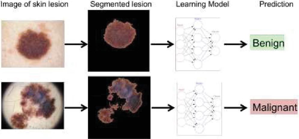
Figure 1 PC vision and AI demonstrative device for specialists and patients to screen dubious skin lesions and moles.
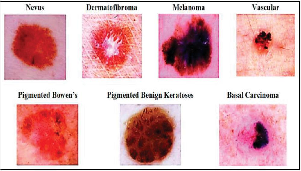
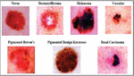
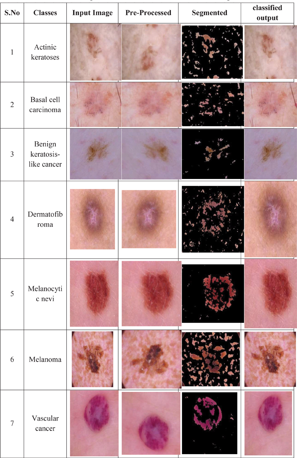
Figure 2 Skinsickness classifications from ISIC dataset.
2 DL Method for Skin Cancer Detection
DNN assume a huge part in skin malignant growth recognition. The majority of the time, a biopsy is required to make a certain cancer diagnosis. Doctors examine cell specimens underneath a microscopy in a laboratory environment. Human cells have a consistent appearance, comparable dimensions, and a well-organized layout. They comprise of a bunch of interconnected hubs. Around 10 billion linked synapses make up the brain. In the subcortical structures, neurons are specialized nerve neurons that receive or transmit messages. The dendrite branch of a cell connects to a hundred nerve cells. Their design is like human cerebrum with regards to neuronal interconnectedness. Their hubs work helpfully to tackle specific issues. Nerve cells, which are cell body expansions that absorb messages from other cells, & axonal, which are cytoplasmic projections that could transmit information to other neurons, are two mechanisms by which neurons acquire associated. The brain as well as spinal cord are encased in three levels of nerves. The pia mater is the fragile innermost lining. The arachnoid, a web-like arrangement containing fluid that safeguards the brain is the middle part. The ground substance is the name of the hard outermost surface. Neural networks are prepared for specific undertakings; in this manner, the networks fill in as specialists in the spaces in which they were prepared. In our review, neural networks were prepared to group pictures and to recognize different kinds of skin disease. Acne, clogged skin pores that let germs, dead skin, as well as oil accumulate in the face. Carcinoma areata, which causes patchy hair loss. Eczema, or contact dermatitis, is characterized by rough, irritated skin that becomes swollen, cracked, or scaly. Research connected with every one of these profound neural networks is examined exhaustively in this part [7]. Shown in Figure 2.
3 (ANN)-Based Skin Cancer Detection Methods
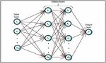
Brain association is a nonlinear as well as verifiable conjecture technique. Its development is procured from normal plan of human brain. Humans need nonlinear synchronisation systems for flow of information across cortical regions during neural impulses, hence variation is crucial. Moderate layers are suggested as concealed layers. In an ordinary ANN, there is couple of mystery layers. The operation of a neural network can be decomposed into particular information operations due to convolutional nodes. Every operation in a hidden layer is tailored to deliver a certain result. Temporary neurons send data to 3rd layer of result neurons. Estimations are learned at every layer utilizing backpropagation, which is utilized for learning convoluted affiliations/associations among data as well as result layers. Backpropagation is a supervised classifier that artificial neural systems employ to calculate a learning algorithm with regard to weighting factor for the multiple parameters. It resembles a brain association [8]. At this point, in programming, the term brain association and fake brain association are used alternately. The brain is a sophisticated organ that manages every bodily function as well as thinking, recollection, empathy, sensation, motor function, sight, respiration, heat, and appetite. The centralized neurological network, or CNS, is made up of the spinal column that emerges from the brain. The fundamental plan of an ANN network is given in Figure 3.
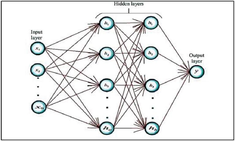
Figure 3 Basic ANN structure.
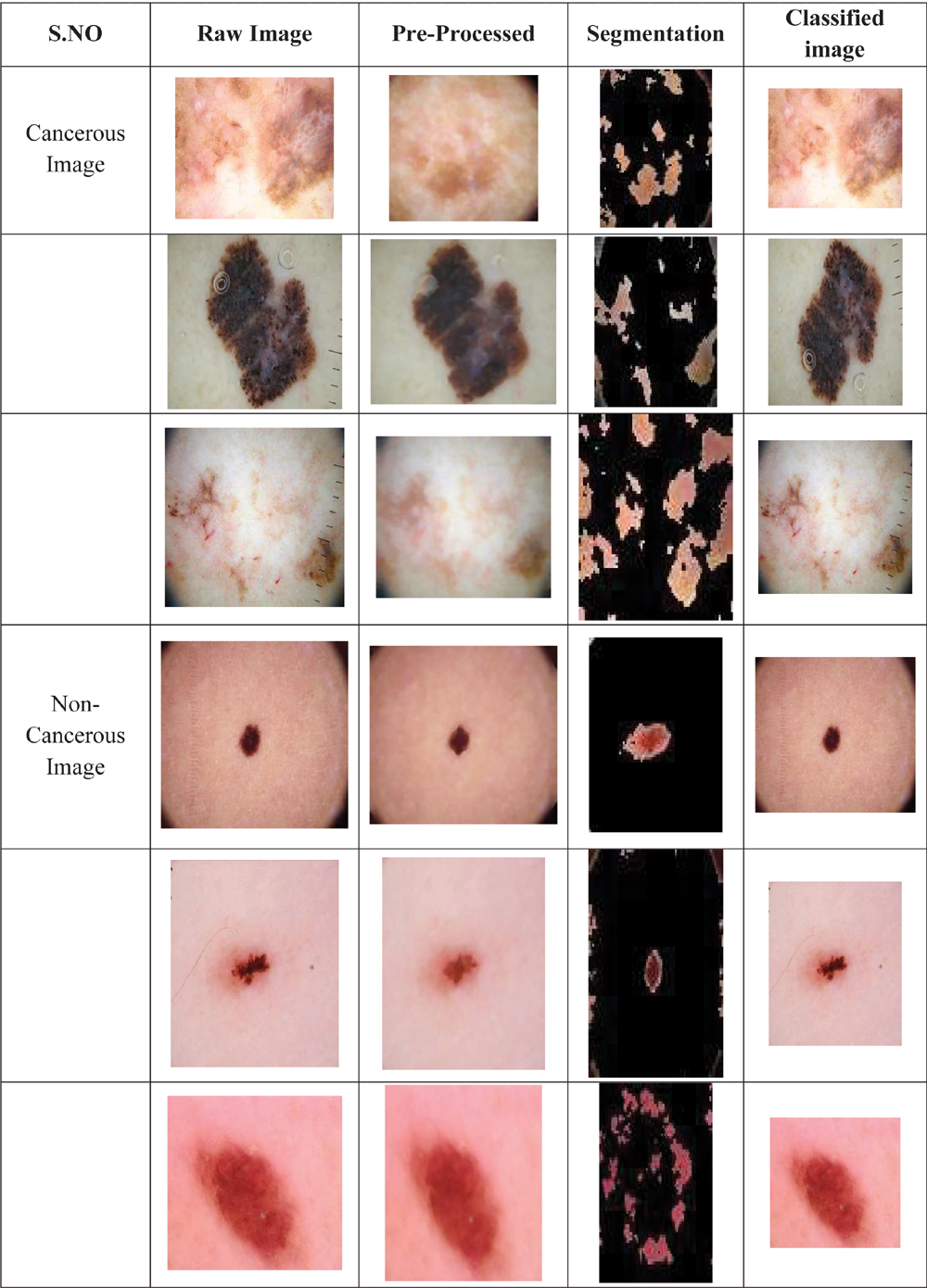
ANN is utilized for request for isolated features in skin threatening development ID structures. Input pictures are designated melanoma or nonmelanoma after successful readiness/course of action of arrangement set. Amount of mystery layers in an ANN depends upon amount of data pictures. The following guideline should be used to decide if or not hidden units are necessary: it is only necessary in artificial neural systems when non-linear data separation is necessary. Data/first layer of ANN cycle partners with mystery layer by data dataset. Dataset is stamped or unlabeled, which is taken care of similarly using an oversaw or solo learning framework. A brain association uses backpropagation or feed-forward designing to learn loads present at every association affiliation/interface. Synaptic weights can be updated using the training algorithm technique to move down that slope. In short, the backward stage of the method determines how significantly the synapse values of every neuronal contributes towards the inaccuracy but then modifies those parameters to enhance the effectiveness of the network. The two models use a substitute model for the underlying dataset. Data streams just from commitment to outcome layer [9]. Continuously transferring information at a fast speed is known as data streaming. Some bitstreams transmit out massive groupings of smaller-sized database files at once while constantly gathering information from countless sources. Work [10] proposed a skin sore portrayal method that arranged injuries into two essential classes: innocuous as well as compromising. Stress fractures or overtraining are the two main categories of accidents. A singular, major catastrophe is typically the cause of acute injuries. Wrist cracks, ankle injuries, elbow displacements, or hamstrings muscle injury are a few typical instances. Proposed method worked in 3 phases. In hidden stage, a self-making NN was utilized to isolate sores from pictures. In ensuing stage, features, for instance, development line, surface, and assortment nuances were extracted. Among the input as well as output levels of a synthetic CNN architecture is a layer known as the hidden layer, wherein perceptron process a collection of input nodes to generate an outcome using an input vector. Author [11] proposed an ANN-based robotized skin illness suggestive structure. Artificial neural systems (ANNs) are computational models with biological influences that mimic how the natural brain processes data. ANNs educate (or are taught) via experiencing rather than by coding, and they learn by identifying trends as well as connections in information. Introduction of three ANN’s learning estimations like LM [12], versatile backpropagation (RP) [13], scaled form slope (SCG) [14], suggested by this research. Relationship of execution showed that LM estimation achieved most imperative identity score (95.1%) and remained capable at gathering of innocuous injuries, while SCG learning computation conveyed superior outcomes if amount of ages was extended, scoring a 92.6% responsiveness regard. A mole portrayal method for early finding of melanoma skin illness was proposed [15, 16]. Processing of cancerous and non-cancerous cell using deep learning technique is shown by Table 1.
Table 1 Processing of cancerous and non-cancerous cell using deep learning techniques
4 Preprocessing Techniques for Skin Lesion Images
Preprocessing is significantly utilized for planning pictures for better handling and precise component identification. Preprocessing incorporates procurement of picture as info, acquiring the grayscale picture, commotion sifting and double picture age [17]. These techniques incorporate differentiation change which plays out the augmentation of histogram of a picture for better perceivability; power change which improves picture’s intensity values to create a result picture with top notch show; and histogram adjustment which appropriates the pixel forces uniformly for the whole scope of powers fully intent on expanding the worldwide difference of pictures. The collection of pixel intensities across a reference line or multi-line pathway in a picture that are taken at periodic intervals to form the concentration profiling of the picture. Navigate to the Properties panel when the Hue/Saturation modification level is chosen in the Settings option to view the parameters for this change. To create all the colours in the image more vibrant, move the Intensity adjuster towards the right. We likewise have binarization which is the most common way of taking a grayscale picture and changing it over completely to high contrast tones simply by diminishing the data; morphological activity which performs disintegration and enlargement on pictures to remove a few elements and area of each and every item in a picture [18]. A grayscale image has pixels with brightness values that vary from 0 to 255. The RGB data (24 bit) of a colour image are converted into grayscale to produce an image representation (8 bit). Grayscale images are created using a variety of image manufacturing techniques and productivity tools. It is significantly simpler to compare colours in grayscale than in RGB. Source code like grayscale over RGB colour space because of this. The method of obtaining characteristics from pictures of analysed lesion patches is known as feature retrieval. A structural component is added by linguistic structures to a source image to generate a result image of the equivalent dimension. When performing a wavelet transforms, every resulting pixel’s value is determined by comparing to it neighbours in the image pixels.
5 Division Procedures for Skin Lesions
Picture division is a significant cycle in robotizing skin lesion conclusion. It is a significant stage in skin lesion pictures examination. This cycle gets locale of interests by isolating infected region from the sound area. This part examines different methods for division of skin lesions. These procedures incorporate handcrafts features based strategies, for example, limit based [19], edge and area-based techniques [20] and insight based administered division. Astute based approaches incorporate artificial neural networks and deep learning strategies [21]. Convolutional Neural Networks are the predominant structure utilized for image identification and detection applications (CNNs). Among the activities that deep neural networks (DNNs) excelled at is enhance recognition. Computer programmes called neural pathways are made to spot similarities. They name themselves from the design of the human brain in the sense of architecture.
AI in light of the elite exhibition picture is utilized to distinguish skin disease that accomplished great proficiency in recognizable proof [22]. Notwithstanding, the exactness of the model can be expanded by removing more highlights and responsiveness is more strayed. In [23], the creator proposed a strategy utilizing picture handling steps that assists with expanding the location exactness of skin malignant growth. Nonetheless, they couldn’t portray a particular model that can effectively identify malignant growth. DL in light of method-driven design is constructed rapidly to such an extent for that reason the model can foresee the outcome as fast. It obtained an improved outcome in identifying of skin disease [24]. Not with standing, the approach calls for continuous interacting with clinical pictures so it can work on the clinical field. In [25], the creator proposed CNN based skin malignant growth recognition where element is separated from dermoscopic pictures utilizing highlight removing strategies. A medical assessment as well as epidermal investigation are usually the first steps in assessing a suspected skin lesion. Dermoscopy, often referred to as dermatoscopy, or interface microscopy, is frequently utilized by dermatology to more thoroughly inspect the disease. Difficulties of skin lesions identification: a: hair relics, b: low differentiation, c: sporadic limits, d: variety enlightenmentan exactness of identification 89.5% in testing stage. In any case, the precision of discovery was not adequate that was expected to get to the next level. In [26], the creator proposed LIN in light of DL to identify and grouping of skin malignant growth. They gained incredible outcome with DL based LIN by separating more highlights. Notwithstanding, division execution was expected to increment for additional upgrading of result [27]. In [28], the creator proposed a DCNN that consolidates three stages that perform phenomenally to distinguish skin lesions where right off the bat, variety change is utilized to improve contrast; furthermore, CNN approach is utilized to extricate lesion limits; at long last, move learning is utilized to separate deep elements. Be that as it may, the strategy accomplished a decent outcome for certain seasons of dataset however results might change for an alternate dataset. In [29], the creator proposed a CNN based method to distinguish melanoma skin malignant growth where they utilized preprocessing as well as post-handling of picture for upgrade when division, separately. The model delivered lesion districts by joining neighborhood and worldwide relevant data. It obtained a decent outcome for the expectation and order. Nonetheless, execution time isn’t referenced which can expand worth of outcomes [30]. Accuracy, review, and F1 score are insufficient high however by making preprocessing strides outcomes might be enhanced with a superior characterization pace of skin lesions pictures.
6 CNN Based Skin Cancer Detection Techniques
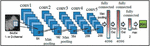
CNN is a fundamental kind of DNN, which is successfully being used in PC vision. It is used for ordering pictures, gathering a gathering of data pictures, and performing picture acknowledgment [31]. Three significant sorts of layers associated with making CNN are convolution layers, pooling layers, and full-associated layers. Fundamental design of a CNN is introduced in Figure 4.
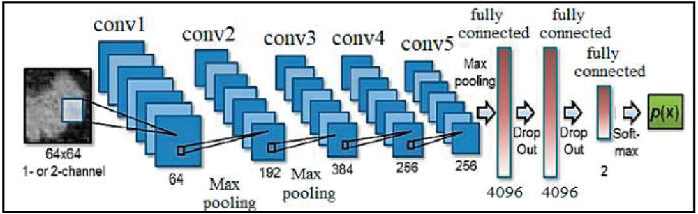
Figure 4 Basic CNN Architecture [9].
CNN-based computerized DL calculations have accomplished astounding execution in identification, division, and arrangement activities of clinical imaging [32]. Creator [33] proposed an exceptionally deep CNN for melanoma discovery. FCRN having 16 remaining blocks was utilized in division cycle to further develop execution. Proposed strategy utilized a normal of both SVM as well as softmax classifier for characterization. It gives 85.5% precision in melanoma grouping with division and 82.8% without division [34]. Coarse-scale was utilized to catch shape attributes by and large relevant data of lesions. Interestingly, the better scale accumulated printed detail of lesion for separation between different kinds of skin lesions. Skin lesions can be categorised by doctors as pre-cancerous tumours (BCC), innocuous or non-malignant (nevi), colored benign keratoses (BKL), squamous cell carcinomas (SCC) as well as carcinogenic (melanoma). Creator [35] proposed a strategy to remove deep elements from different well established and pre-prepared deep CNNs for skin lesions order. A suspect region will be excised and taken to a laboratory to be examined underneath a magnification if the help diagnose it may be a cancer. It is known as a skin biopsy. At last, the classifier results were intertwined to perform characterization [36]. At first, it was prepared on 3797 lesion pictures; be that as it may, later, 29-times expansion was applied in view of lighting positions and scale changes. A method for grouping of four unique kinds of skin lesion pictures was proposed by [37]. Every region of skin that differs in colour, form, dimension, or thickness from the outer tissue is referred to as a skin lesion. It is relatively prevalent and frequently develop from localised skin irritation, such as skin infections or sun exposure.
Work [38] proposed a KNN-based skin infection area method. Dimensionality of assessed spectra was diminished with PCA procedure. Both KNN as well as ANN were ready as well as their show for melanoma area was checked out. On test dataset, request error of KNN was 2–3%, while gathering bumble for ANN lay in extent of 3% to 4%. Proposed structure eliminated assortment, GLCM, and morphological components of injury pictures, after which course of action method included those features as data. Also, gathering execution of proposed structure was differentiated and KNN, ANN, and guileless Bayes classifiers. Proposed method attained 93.150685% accuracy while KNN 71.232877%, ANN 63.013699%, and gullible Bayes 56.164384% precision scores. Another KNN motorized skin dangerous development decisive system was proposed by [38]. Proposed system used a center channel as an uproar clearing method. Then, filtered pictures were segmented with a real region creating as well as uniting strategy. Verifiable components were taken out from injury pictures, while printed features were isolated from a curvelet space. Finally, proposed method described data pictures into unsafe or noncancerous with 98.3% accuracy. In this research, various classifiers like SVM, BPN, and 3-layer NN were moreover executed, and their display was differentiated and proposed structure’s portrayal execution. SVM made 91.1% accuracy, BPN 90.4% precision, 3-layer NN 90.5%, however the proposed structure attained most significant accuracy of 98.3% for skin infection finding.
Work [39] proposed a GAN-based skin sore request method. Proposed structure performed increment on a planning set of pictures with reasonable looking skin injury pictures delivered through GAN. CNN learned to arrange seven exceptional classes of skin sores. Results of proposed method were differentiated and ResNet-50 and DenseNet. ResNet-50 conveyed 79.2% precision. Work [40] suggested a GAN-based skin sore gathering method. Proposed structure performed development on a readiness set of pictures with sensible looking skin sore pictures made through GAN. CNN sorted out some way to bunch seven interesting classes of skin injuries. Delayed consequences of proposed method were differentiated and ResNet-50 and DenseNet. ResNet-50 made 79.2% accuracy, DenseNet showed 81.5% precision, however proposed method achieved most raised accuracy of 86.1% for skin sore request. Profound learning procedures give satisfactory accuracy yet require pure, unbalanced, and colossal arrangement datasets. To overcome these limitations, [41] proposed a profound learning method for data cleaning as well as GAN for data increment. Proposed method utilized decoupled profound convolutional GANs for data age. Proposed structure beat benchmark ResNet-50 method for skin sore portrayal and achieved 86.1% precision. A smart data increment technique for a skin injury in light of self-thought moderate PGAN was proposed. Moreover, generative method was improved with change methodology. Proposed structure attained 70.1% accuracy as differentiated and 67.3% precision made by a non-extended method. Work [42] proposed a method to recognize period of melanoma threatening development considering disease thickness. Move learning with VGG16 CNN is utilized. This method bunches melanoma in 3 stages with 87.2% precision. Maker [43] proposed a system considering the hard and fast dermoscopic score to perceive period of melanoma. Picture is preprocessed utilizing rgb2gray change and center channel to decrease upheaval. Sobel edge recognizing verification computation is utilized. Lopsidedness, Border peculiarity, Color assortment, Differential plan are features utilized to figure hard and fast dermoscopic score. Work [44] proposed a structure which orders developments into two classes considering thickness of melanoma as slim as well as thick melanomas. High level dermoscopy assessment uses motorized examination of electronic pictures and it deals assessment of morphological components of sores by solidification with the item. Gong et al. (2020) proposed a decision blend system which relies upon various pre-arranged CNN. This essentially handles theory issue of CNN. StyleGANs are arranged utilizing ISIC 2019 dataset as well as this further fosters gathering accuracy. Maker [45] suggests a bi-directional dermoscopic incorporate learning plan which concentrates features. Picture parsing limit is further developed by controlling part multiplication. Work [46] proposed acknowledgment and division of skin dangerous development from dermoscopic pictures. It relies upon the lightweight profound learning organization. This strategy can eliminate discriminative sore features as well as moreover further fosters the ID execution of the model. Work [47] proposed scheme that usages further created whale improvement estimation to propel CNN. This better estimation is utilized for best assurance of burdens as well as inclinations in organization to diminish bungle of the organization result as well as best outcome. The various stages of skin cancer processing is shown in Table 2. Work [48] developed an automated and computerised system for spotting skin conditions has been developed using deep learning techniques. The performance of various neural network methods is assessed and validated in the proposed system to detect & detect skin disorders across a variety of parameters. Work [49] suggested the models produced using data augmentation techniques outperform those created by starting from scratch. Results demonstrate that image enhancement strategies had a considerable impact on the systems both prior to and after using methods. Work [50] proposed the programs for e-healthcare that are Internet of Things (IoT) capable are helping the community more by offering medical monitoring necessary assistance in intelligent environments. Considerations involving medical data privacy as well as safety arise when personal medical information is stored safely in a digital form.
Table 2 Processing of various skin cancer classes utilizing DL methods
7 Challenge of Skin Cancer Detection
A few troubles to identify skin malignant growth that can be credited to variety in picture types and sources. The variety in presence of human skin variety makes skin malignant growth discovery harder as well as complex. However, this prevalent type of carcinoma can also develop on parts of the body that are not often sensitive to sunshine. Basal cell carcinoma, melanoma, besides squamous cell carcinoma are the three main kinds of skin cancer. These difficulties and most visual attributes of skin lesions pictures are depicted beneath:
1. Principal hardships in skin malignant growth are few sizes and states of pictures that can’t give a precise consequence of ID. In this viewpoint, pre-handling is expected for exact examination.
2. A couple of squandered signals are to be compromised that are not initially a piece of a picture however can hinder to obtain a decent outcome. In this way, in pre-handling steps, this large number of commotion and curios ought to be taken out.
3. Now and again, low differentiation from adjoining tissues represents extra troubles and makes it harder to unequivocally dissect skin disease.
4. Variety enlightenment additionally makes a few troubles having its elements like variety surface, light beams, and reflections.
5. Human body exists a few moles which might in all likelihood never foster disease cell yet they make a few challenges to identify skin malignant growth precisely from harmful pictures.
6. The ongoing inclination that contorts the exhibition of the models to accomplish an improved outcome is another test in regards to skin disease recognition.
The principal purpose of this review was to foster different DL methods for arrangement of tumour skin disease as well as then to analyze presentation consequences of these models. We adjusted a few DL designs. Image Data Generator innovation was utilized to defeat the information volume issue. The initial data sources are utilized to feed the Keras Image Data Generator, which transforms the information randomly and outputs an output that only contains the freshly altered information. We then, at that point, assessed and looked at the model exhibition for the designs that were all thought of, in light of normal execution measures. While contrasting this review and past observational investigations, for example, [48], we observed that the differentiation of this study is that we utilized a few CNN structures. The outcomes contrasted among consequences of this study might be great, somewhat. We may likewise be more far reaching in adjusting a few models, as the outcomes might be enhanced by utilizing a bigger dataset. Some exploration studies [49] have shown gigantic information to prepare the network well and, in this way, give more precise outcomes. Be that as it may, one of the difficulties related with this is the accessibility of excellent equipment, like GPUs. For instance, our examination on a humbly estimated dataset (7146 pictures) required around six and a half hours to play out all tasks with eight models, despite the fact that we utilized GPUs utilizing Colaboratory. Bigger dataset, additional time that should be spent. A few examinations [50] have demonstrated that CNNs ought to be prepared on excellent information, as loud picture information might increment misclassification rates. We saw, over the span of the trial, that a few pictures contained some clamor, which might have diminished their excellence. Subsequently, the mining of fundamental highlights might be impacted while preparing some unacceptable preparation on certain elements. Subsequently, guaranteeing just great pictures are accessible in the dataset is a basic matter.
8 Discussion
Here utilized a reasonable measure of the preparation and assessment information in the examination. Accordingly, we plan to expand upon this trial using bigger picture datasets, as a few investigations have demonstrated that convolutional neural networks ought to be taken care of broad information to get results with exceptionally high precision. Through aftereffects of this review, we planned to foster mechanical frameworks for use in day-to-day existence and to help dermatologists in really distinguishing skin malignant growth. In addition, the more pictures of melanoma of various sorts that CNN is presented to, higher its exactness in perceiving various elements. This subject might be fascinating in future exploration. Nonetheless, dissimilar to past investigations, we played out a definite exploratory concentrate by embracing a scope of deep learning designs to relatively evaluate their presentation conduct and to recognize the skin malignant growth utilizing clinical pictures. As every DL engineering had a different number of handling layers, it very well may be seen. Deep learning (DL) requires equipment assets, for example, GPU to accelerate the preparation time. Not at all like existing examinations, we included late DL designs utilizing the ISIC benchmark dataset to recognize the proper DL engineering while explicitly handling the melanoma skin disease grouping task. Significant preparation was performed on a start to finish premise utilizing melanoma and non-melanoma skin malignant growth pictures. Consequently, we show attainability of DL while finding regardless of whether skin has melanoma without requirement for area master’s mediation for component designing part. We believe that it is critical to decide how to execute these outcomes socially after framework improvement and interface them to clients’ advantage. Contingent upon the idea of framework advancement, the endpoint and the kind of picture information expected for the improvement will change [51]. For instance, on the off chance that the individual who utilizes the framework is a specialist, profoundly exact framework improvement more like an affirmed conclusion will be needed. Preparing NNs that can recognize malignant growths from dermoscopic pictures will be additionally out of luck. Notwithstanding, for in-clinic use there is now a demonstrative strategy: biopsy. Biopsy is a strategy for partaking in skin tissue as well as making a neurotic finding. Through a biopsy, it is feasible to make a practically 100 percent finding (affirmed determination). Also, the strategy of biopsy takes something like 10 min. Then again, while considering their utilization by the overall population outside clinical organizations, it is challenging to exhibit their demonstrative presentation completely. This is on the grounds that the reproducibility of shooting conditions can’t be guaranteed, and shooting gear is unique. Consequently, while utilizing an imaging framework outside clinical organizations, it could be smarter to utilize the framework to point out skin disease instead of spotlight on working on demonstrative execution. Additionally, nobody can say that the exactness of the framework should be improved when it is utilized external the clinical establishment.
9 Conclusion
This survey is a basic and scientific overview of the cutting edge techniques for performing investigation of skin lesion pictures. Melanoma skin malignant growth is exceptionally hazardous and gets metastasized extremely quick. It is expected to recognize the phase of malignant growth to begin the conclusion. Characterization of the phases of dermoscopic pictures is viewed as one of the difficult errands. Grouping of the phases of malignant growth is an exceptionally dreary undertaking and vital when patient conclusion is thought of. This paper proposed harmless stage grouping arrangement of melanoma skin disease. Two frameworks are presented in this; two phase arrangement situation which, right off the bat, orders melanoma in 3 dissimilar stages. Here used appraisal plan from melanoma-10-overlap dataset to survey our calculation, contrasted and ground truth, and figured a few boundaries like particularity, responsiveness, exactness, MSE, RMSE and RSE. We exhibited system achieved a higher precision, particularity and responsiveness than existing structures as well as moreover it has lower level of RAE as well as RRES contrasted with other comparable strategies (misfortune capabilities). The proposed technique is contrasted and SVM and CNN, it gives superior implementation.
Declarations
Funding
No funds, grants were received by any of the authors.
Conflict of interest
There is no conflict of interest among the authors.
Data Availability
All data generated or analysed during this study are included in the manuscript.
Code Availability
Not applicable.
Author’s Contributions
All author is contributed to the design and methodology of this study, the assessment of the outcomes and the writing of the manuscript.
References
[1] Rezayi, S.; Mohammadzadeh, N.; Bouraghi, H.; Saeedi, S.; Mohammadpour, A. Timely Diagnosis of Acute Lymphoblastic Leukemia Using Artificial Intelligence- Oriented Deep Learning Methods. Comput. Intell. Neurosci. 2021, 2021, 5478157.
[2] Tufail, A.B.; Ma, Y.K.; Kaabar, M.K.; Martínez, F.; Junejo, A.; Ullah, I.; Khan, R. Deep learning in cancer diagnosis and prognosis prediction: A minireview on challenges, recent trends, and future directions. Comput. Math. Methods Med. 2021, 2021, 9025470.
[3] Li, X.; Jiao, H.; Wang, Y. Edge detection algorithm of cancer image based on deep learning. Bioengineered 2020, 11, 693–707
[4] Alzubaidi, L.; Zhang, J.; Humaidi, A.J.; Al-Dujaili, A.; Duan, Y.; Al-Shamma, O.; Santamaría, J.; Fadhel, M.A.; Al-Amidie, M.; Farhan, L. Review of deep learning: Concepts, CNN architectures, challenges, applications, future directions. J. Big Data 2021, 8, 1–74.
[5] Ghods, A.; Cook, D.J. A survey of deep network techniques all classifiers can adopt. Data Min. Knowl. Discov. 2021, 35, 46–87.
[6] Emanuelli, M.; Sartini, D.; Molinelli, E.; Campagna, R.; Pozzi, V.; Salvolini, E.; Simonetti, O.; Campanati, A.; Offidani, A. The double-edged sword of oxidative stress in skin damage and melanoma: From physiopathology to therapeutical approaches. Antioxidants 2022, 11, 612.
[7] Ferlay, J.; Colombet, M.; Soerjomataram, I.; Parkin, D.M.; Piñeros, M.; Znaor, A.; Bray, F. Cancer statistics for the year 2020: An overview. Int. J. Cancer 2021, 149, 778–789.
[8] Sung, H.; Ferlay, J.; Siegel, R.L.; Laversanne, M.; Soerjomataram, I.; Jemal, A.; Bray, F. Global cancer statistics 2020: GLOBOCAN estimates of incidence and mortality worldwide for 36 cancers in 185 countries. CA Cancer J. Clin. 2021, 71, 209–249.
[9] Abdar, M.; Samami, M.; Mahmoodabad, S.D.; Doan, T.; Mazoure, B.; Hashemifesharaki, R.; Liu, L.; Khosravi, A.; Acharya, U.R.; Makarenkov, V.; et al. Uncertainty quantification in skin cancer classification using three-way decision- based Bayesian deep learning. Comput. Biol. Med. 2021, 135, 104418.
[10] Hosny, K.M.; Kassem, M.A.; Foaud, M.M. Classification of skin lesions using transfer learning and augmentation with Alex-net. PLoS ONE 2019, 14, e0217293.
[11] Khamparia, A.; Singh, P.K.; Rani, P.; Samanta, D.; Khanna, A.; Bhushan, B. An internet of health things-driven deep learning framework for detection and classification of skin cancer using transfer learning. Trans. Emerg. Telecommun. Technol. 2021, 32, e3963.
[12] Nahata, H.; Singh, S.P. Deep learning solutions for skin cancer detection and diagnosis. In Machine Learning with Health Care Perspective; Springer: Cham, Switzerland, 2020; pp. 159–182.
[13] Demir, A.; Yilmaz, F.; Kose, O. Early detection of skin cancer using deep learning architectures: Resnet-101 and inception-v3. In Proceedings of the 2019 Medical Technologies Congress (TIPTEKNO), Izmir, Turkey, 3–5 October 2019; pp. 1–4.
[14] Cassidy, B.; Kendrick, C.; Brodzicki, A.; Jaworek-Korjakowska, J.; Yap, M.H. Analysis of the ISIC image datasets: Usage, benchmarks and recommendations. Med. Image Anal. 2022, 75, 102305.
[15] Abbas, Q.; Ramzan, F.; Ghani, M.U. Acral melanoma detection using dermoscopic images and convolutional neural networks. Vis. Comput. Ind. Biomed. Art 2021, 4, 25.
[16] Reethu, R.; Preetha, D.; Parameshwaran, P.; Sivaparthipan, C. B.; Kalaikumaran, T. A design of smart device for detection of oral cancer using IoT. Int J Res Eng Sci Manag 2020, 3(3), 44–47.
[17] Sayed, G.I.; Soliman, M.M.; Hassanien, A.E. A novel melanoma prediction model for imbalanced data using optimized SqueezeNet by bald eagle search optimization. Comput. Biol. Med. 2021, 136, 104712.
[18] Mijwil, M.M. Skin cancer disease images classification using deep learning solutions. Multimed Tools Appl. 2021, 80, 26255–26271.
[19] Nawaz, M.; Mehmood, Z.; Nazir, T.; Naqvi, R.A.; Rehman, A.; Iqbal, M.; Saba, T. Skin cancer detection from dermoscopic images using deep learning and fuzzy k- means clustering. Microsc. Res. Tech. 2022, 85, 339–351.
[20] Dorj, U.; Lee Ke Choi, J.; Lee, M. The skin cancer classification using deep convolutional neural network. Multimed. Tools Appl. 2018, 77, 9909–9924.
[21] Afza, F.; Sharif, M.; Mittal, M.; Khan, M.A.; Jude Hemanth, D. A hierarchical three- stepsuperpixels and deep learning framework for skin lesion classification. Methods 2022, 202, 88–102.
[22] Hameed, N.; Shabut, A.M.; Ghosh, M.K.; Hossain, M. Multi-class multi-level classification algorithm for skin lesions classification using machine learning techniques. Expert Syst. Appl. 2020, 141, 112961.
[23] Singh, L.; Janghel, R.R.; Sahu, S.P. TrCSVM: A novel approach for the classification of melanoma skin cancer using transfer learning. Data Technol. Appl. 2021, 55, 64–81.
[24] Arshad, M.; Khan, M.A.; Tariq, U.; Armghan, A.; Alenezi, F.; Javed, M.Y.; Aslam, S.M.; Kadry, S. A computer-aided diagnosis system using deep learning for multiclass skin lesion classification. Comput. Intell. Neurosci. 2021, 2021, 9619079
[25] Khan, M.A.; Akram, T.; Zhang, Y.-D.; Sharif, M. Attributes based skin lesion detection and recognition: A mask RCNN and transfer learning-based deep learning framework. Pattern Recognit. Lett. 2021, 143, 58–66.
[26] Abunadi, I.; Senan, E.M. Deep learning and machine learning techniques of diagnosis dermoscopy images for early detection of skin diseases. Electronics 2021, 10, 3158.
[27] Naeem, A.; Farooq, M.S.; Khelifi, A.; Abid, A. Malignant melanoma classification using deep learning: Datasets, performance measurements, challenges and opportunities. IEEE Access 2020, 8, 110575–110597.
[28] Malik, H.; Anees, T. BDCNet: Multi-classification convolutional neural network model for classification of COVID-19, pneumonia, and lung cancer from chest radiographs. Multimed. Syst. 2022, 28, 815–829
[29] Naeem, A.; Anees, T.; Naqvi, R.A.; Loh, W.-K. A comprehensive analysis of recent deep and federated-learning-based methodologies for brain tumor diagnosis. J. Pers. Med. 2022, 12, 275.
[30] Deeba, F.; Kun, S.; Dharejo, F.A.; Zhou, Y. Sparse representation based computed tomography images reconstruction by coupled dictionary learning algorithm. IET Image Process. 2020, 14, 2365–2375.
[31] Zawish, M.; Siyal, A.A.; Ahmed, K.; Khalil, A.; Memon, S. Brain tumor segmentation in MRI images using Chan-Vese technique in MATLAB. In Proceedings of the 2018 International Conference on Computing, Electronic and Electrical Engineering (ICE Cube), Quetta, Pakistan, 12–13 November 2018; pp. 1–6.
[32] Eraslan, G.; Avsec, Ž.;Gagneur, J.; Theis, F.J. Deep learning: New computational modelling techniques for genomics. Nat. Rev. Genet. 2019, 20, 389–403.
[33] Khan, M.A.; Muhammad, K.; Sharif, M.; Akram, T.; Kadry, S. Intelligent fusion- assisted skin lesion localization and classification for smart healthcare. Neural Comput. Appl. 2021, 1–16.
[34] Chaturvedi, S.S.; Tembhurne, J.V.; Diwan, T. A multi-class skin cancer classification using deep convolutional neural networks. Multimed. Tools Appl. 2020, 79, 28477–28498.
[35] Deeba, F.; Kun, S.; Dharejo, F.A.; Zhou, Y. Wavelet-based enhanced medical image super resolution. IEEE Access 2020, 8, 37035–37044.
[36] Dharejo, F.A.; Deeba, F.; Zhou, Y.; Das, B.; Jatoi, M.A.; Zawish, M.; Du, Y.; Wang, X. TWIST-GAN: Towards wavelet transform and transferred GAN for spatio- temporal single image super resolution. ACM Trans. Intell. Syst. Technol. (TIST) 2021, 12, 1–20
[37] Jeny, A.A.; Sakib, A.N.M.; Junayed, M.S.; Lima, K.A.; Ahmed, I.; Islam, B. SkNet: A convolutional neural networks based classification approach for skin cancer classes. In Proceedings of the 2020 23rd International Conference on Computer and Information Technology (ICCIT), Dhaka, Bangladesh, 19–21 December 2020; pp. 1–6.
[38] AAli, S.; Miah, S.; Haque, J.; Rahman, M.; Islam, K. An enhanced technique of skin cancer classification using deep convolutional neural network with transfer learning models. Mach. Learn. Appl. 2021, 5, 100036.
[39] You, Y.; Zhang, Z.; Hsieh, C.; Demmel, J.; Keutzer, K. Imagenet training in minutes. In Proceedings of the 47th International Conference on Parallel Processing, Eugene, OR, USA, 13–16 August 2018; pp. 1–10.
[40] Quang, N.H. Automatic skin lesion analysis towards melanoma detection. In Proceedings of the 2017 21st Asia Pacific Symposium on Intelligent and Evolutionary Systems (IES), Hanoi, Vietnam, 15–17 November 2017; pp. 106–111.
[41] Amin, J.; Sharif, A.; Gul, N.; Anjum, M.A.; Nisar, M.W.; Azam, F.; Bukhari, S.A.C. Integrated design of deep features fusion for localization and classification of skin cancer. Pattern Recognit. Lett. 2020, 131, 63–70.
[42] Aburaed, N.; Panthakkan, A.; Al-Saad, M.; Amin, S.A.; Mansoor, W. Deep convolutional neural network (DCNN) for skin cancer classification. In Proceedings of the 2020 27th IEEE International Conference on Electronics, Circuits and Systems (ICECS), Glasgow, UK, 23–25 November 2020; pp. 1–4.
[43] Zhang, J.; Xie, Y.; Xia, Y.; Shen, C. Attention residual learning for skin lesion classification. IEEE Trans. Med. Imaging 2019, 38, 2092–2103.
[44] Liu, L.; Mou, L.; Zhu, X.X.; Mandal, M. Automatic skin lesion classification based on mid-level feature learning. Comput. Med. Imaging Graph. 2020, 84, 101765.
[45] Höhn, J.; Hekler, A.; Krieghoff-Henning, E.; Kather, J.N.; Utikal, J.S.; Meier, F.; Gellrich, F.F.; Hauschild, A.; French, L.; Schlager, J.G.; et al. Integrating patient data into skin cancer classification using convolutional neural networks: Systematic review. J. Med. Internet Res. 2021, 23, e20708.
[46] Esteva, A.; Kuprel, B.; Novoa, R.A.; Ko, J.; Swetter, S.M.; Blau, H.M.; Thrun, S. Dermatologist-level classification of skin cancer with deep neural networks. Nature 2017, 542, 115–118.
[47] Maron, R.C.; Weichenthal, M.; Utikal, J.S.; Hekler, A.; Berking, C.; Hauschild, A.; Enk, A.H.; Haferkamp, S.; Klode, J.; Schadendorf, D.; et al. Systematic outperformance of 112 dermatologists in multiclass skin cancer image classification by convolutional neural networks. Eur. J. Cancer 2019, 119, 57–65.
[48] Hosny, K.M.; Kassem, M.A.; Fouad, M.M. Classification of skin lesions into seven classes using transfer learning with AlexNet. J. Digit. Imaging 2020, 33, 1325–1334.
[49] Chassagnon, G.; Vakalopolou, M.; Paragios, N.; Revel, M.P. Deep learning: Definition and perspectives for thoracic imaging. Eur. Radiol. 2020, 30, 2021–2030.
[50] Acosta, M.F.J.; Tovar, L.Y.C.; Garcia-Zapirain, M.B.; Percybrooks, W.S. Melanoma diagnosis using deep learning techniques on dermatoscopic images. BMC Med. Imaging 2021, 21, 6.
[51] Brinker, T.J.; Hekler, A.; Enk, A.H.; Berking, C.; Haferkamp, S.; Hauschild, A.; Weichenthal, M.; Klode, J.; Schadendorf, D.; Holland-Letz, T.; et al. Deep neural networks are superior to dermatologists in melanoma imageclassification. Eur. J. Cancer 2019, 119, 11–17.
Biographies

CH. Srilakshmi is a Research Scholar in the Department of CSE at JNTU Kakinada, Andhra Pradesh, India. Her research interests may include Deep learning, Robotics, AI, Internet of Things (IoT), Wireless Communications and Underwater Localization.

E. Laxmi Lydia is currently a professor of computer science engineering with the GMRIT. She is also a Big Data Analytics Online Trainer with the International Training Organization. She has presented various webinars on big data analytics. She is with the Government DST Funded Project. She is certified by the Microsoft Certified Solution Developer (MCSD). She is the author of the Big Data Analytics Book. She holds a patent. She has published ten research papers in international conference proceedings. She has published more than 100 research articles in international journals in Big Data Analytics and Data Science.

N. Ramakrishnaiah is currently working as Professor in Computer science and Engineering Department at JNTU Kakinada. Under his guidance 1 scholar was awarded Ph.D and 15 research scholars are working. He published 20+ papers in various reputed international journals and conferences. His areas of research interest are Wireless Networks and Machine Learning.
Journal of Mobile Multimedia, Vol. 19_6, 1583–1606.
doi: 10.13052/jmm1550-4646.19610
© 2023 River Publishers